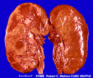
DEFINITION GENERALITY
Amyloidosis was first described in the 19th Century.
Amyloidosis is now known to be a group of diseases in which one or more organ systems in the body accumulates protein deposits.
abnormal protein that may be deposited in any of your body's tissues or organs.
This abnormal protein comes from cells in the bone marrow.
disabling or life threatening.
The disease known as amyloidosis (pronounced am-i-loy-do'-sis) results when enough amyloid protein builds up in one or more organs to cause the organ(s) to malfunction.
 The heart,
  kidneys,
  nervous system
  spleen
 adrenal
 gastro-intestinal tract are most often affected.
the site of deposition depend on the type of amyloidosis primary or secondary.
 Amyloidosis is a bone marrow disease.
 bone marrow makes protective antibodies.
  After they have served their function, these antibodies are broken down and recycled by the system.
 In the amyloidosis, cells in the bone marrow produce antibodies that cannot be broken down.
 These antibodies then begin to build up in the bloodstream.
 Ultimately, they leave the bloodstream and can deposit in your tissues as amyloid.
 amyloid is a hyalin substance that stains specifically with congo red, when examined with ME is found to be composed of fibers lacking periodicity [see specificity chapter]
 amyloid is produced by the reticulum endothelial system and is deposited extracellularily.
recognition in 3 categories:
 typical amyloidosis
 slpeen
 kidney
 liver
liver adrenal
 intestinal mucosa [rectal bipsy for diag]
 atypical amyloidosis
 any organ in addition to those mentioned [amyloidosis of the age affecting more the heart , 3% over 70 years old]
 tumor forming amyloidosis
 nodules of infiltrates by plasma cells may occurs [tongue]
 tumor like deposits [pancreas islets and c cell thyroid tumors [calcitonin producing cells]
There are three major etiologic types, all very different from each other.
1. Primary Amyloidosis
familial mediterranean fever = typical amyloidosis
neuropathic amyloidosis = atypical amyloidosis
Primary amyloidosis is a plasma cell disorder and occasionally occurs with multiple myeloma.
This is the most common type of amyloidosis in the United States and is usually treated (conventionally) with chemotherapy.
Primary amyloid is not associated with any other diseases.
In primary amyloidosis, the organs most often involved include:
 the heart, kidneys,
  nervous system,
  gastrointestinal tract.
 Amyloid deposits in these organs cause:
 shortness of breath,
 fatigue,
  edema (swelling of ankles and legs),
  dizziness upon standing,
 a feeling of fullness in the stomach (especially after eating),
 diarrhea,
  weight loss,
  enlarged tongue,
 numbness of the legs and arms,
  and protein in the urine.
2. Secondary Amyloidosis
Secondary amyloid is caused by a chronic infection or inflammatory disease.
tuberculosis is present in 50% of the cases of amyloidosis
osteomyelitis 12%
chronic lung infection 10%
other chronic infections 12% = hyperimmunization
primary chronic polyarthritis 20% of cases show amyloidosis
Treatment of the underlying chronic infection or inflammatory disease can slow down or stop the progression of this type of amyloid.
In secondary amyloidosis, symptoms caused by the underlying chronic infection or inflammatory disease are complicated by the development of amyloid deposits in the kidney.
This may cause protein in the urine, edema, and fatigue
3. Hereditary Amyloidosis
Familial amyloid is the only type of amyloidosis that is inherited.
It is rare and is found in families of nearly every ethnic background.
In hereditary amyloidosis, the nervous system and gastrointestinal tract are often involved.
This can cause numbness and tingling in the arms and legs, dizziness upon standing, and diarrhea.
Families have their own pattern of organ involvement and symptoms.
The transmission is autosomal dominant, which means that if you have this type of amyloidosis each of your children has a 50% chance of inheriting the disease.
If your child does not inherit the gene, he/she cannot pass it on to future generations.
4. Other types of amyloidosis
include localized amyloid, b2 microglobulin amyloid, and Alzheimer's disease.
Localized types of amyloidosis are associated with hormones, proteins, aging, and specific areas of the body, and are not found with systemic involvment.
The type of amyloidosis which is due to the b2 microglobulin protein may affect people who have been on dialysis for a significant length of time.
In Alzheimer's disease, the amyloid protein in the brain is called the b-amyloid protein.
Amyloidosis can only be diagnosed by a positive biopsy; that is, an identification of the amyloid deposits in a piece of tissue.
 Amyloidosis is rare, affecting about 8 persons per million annually. Its cause is not known.
  It can affect anyone, but the majority of people who get amyloidosis are over the age of 40.
  Primary amyloidosis is not contagious or inherited.
 It is not known how many people have this disease.
  About 10 percent of patients who have multiple myeloma (a form of bone marrow cancer), develop amyloidosis.
  Although amyloid is an abnormal protein, diet and the amount of protein you eat play no role in the development of the disease.
  Also, there is no recognized link between amyloidosis and stress or occupation.
ACQUIRED AMYLOIDOSIS
 Protein
  AL: Immunoglobulin light chains; k or l
 Clinical
  Most common in middle aged males
  Associated disorders
  Paraproteinemia (M-protein)
  In 90% when serum & urine tested by immunofixation
  Most common in nephrotic syndrome
  Least common in polyneuropathy
  Light chain in 1/3
  Intact immunoglobulin in 2/3
  l light chains: AL type
  k light chains: Multiple myeloma; MGUS
  Multiple myeloma : May present 10-81 months after diagnosis of AL
  Polyneuropathy: Occurs in 20% of patients with light chain amyloid
  Distal; Symmetric
  Predominantly sensory
  Pain & temperature loss most prominent
 Distal weakness may develop later
  Autonomic
  Orthostatic hypotension
  Hypoactive pupils
  Hypohydrosis
  Bladder dysfunction
  Impotence
  Carpal tunnel syndrome in 25%
  Myopathy
  Systemic features
  Purpura: Periorbital
  Submandibular swelling
  Cardiac
  Renal: Nephrotic syndrome
  Muscle enlargement ± weakness
  Gastrointestinal: Diarrhea
  Anemia associated with multiple myeloma
  Amyloidoma
  Laboratory
  CSF: Protein mildly elevated
  Electrodiagnostic: Axonal neuropathy
  Biopsy: Axonal loss; Amyloid
  Prognosis
  Gradual progression
  Survival of 1 to 10 years depending on organ involvement
  Shortest survival with cardiac (< 1 year) & renal dysfunction

 HEREDITARY PNS AMYLOIDOSIS
 Mutations in serum proteins that can form b-pleated sheets
 Protein forming the amyloid is identified by immunocytochemistry
 Liver transplantation may be effective therapy
 ATTR: Transthyretin (Prealbumin)
  l Chromosome 18q11.2-q12.1; Dominant
  ATTR gene
  4 exons: Exon 1 codes for signal peptide
  Mutations
  General locations
  Exon 1: Only 1 mutation (Val20Ile); Not in signal peptide
  > 70 in other 3 exons
  Specific mutations: Variable clinical presentation
  Val30Met: Most common; Onset 25 to 65 years; Small fiber & autonomic D
  Val28Met: 7th decade onset neuropathy with impotence; No FH
  Database: HGMD
  Types: Most are missense point mutations
  ATTR Protein
  127 amino acids
  Present in plasma
  Binds: Thyroxine (20%); Retinol binding protein
  Clinical syndromes
  Heterogeneous, with some relation to:
  Point mutation & Ethnic background
  Signs include:
  Peripheral nerve
  Polyneuropathy: Sensory, Autonomic, ± Motor
  Carpal tunnel syndrome
  Systemic: Cardiomyopathy, Vitreous opacities, Renal failure
  Rare CNS
  Glycine 18: Meningocerebrovascular syndrome sparing eyes
  Glycine 30: Oculoleptomeningeal syndrome
  Ile107Val: Autonomic; Carpal tunnel; CNS; Multisystem (Cardiomyopathy; Lung; Joint; Angiopathy)
  General signs: Hearing loss; Migraine; Dementia; Cerebellar; Seizures; Stroke; Myelopathy
  Onset 17-78 years
  Earlier: Portuguese; Japanese endemic Val30Met foci
  Later: Swedish; Sporadic Japanese Val30Met patients
  ? Anticipation: Not explained by triplet repeats
  Homozygotes & heterzygotes with same clinical manifestations
  Penetrance: Variable: 30% to 95%
  FAP I
FAP I  Presentation: Generalized Autonomic & Sensory Polyneuropathy
  Onset: Legs
  Painful dysesthesias
  Most common point mutation: Val30Met
  Onset: 20 to 80 years
  Later: Sporadic Japanese cases; Swedish endemic foci
  Earlier: Japanese & Portugese endemic foci
  Penetrance
  High: Endemic Japanese foci
  Low: Scattered, sporadic cases; Swedish endemic foci
  Male > Female: Especially in late onset sporadic cases
  Weakness: Distal; Legs > Arms
  Sensory loss
  Early onset: Pain & Temperature loss most prominent
  Late onset: Distal; Symmetric; All modalities; Paresthesias
  Tendon reflexes: Reduced or Absent
  Autonomic failure
  Severe in early onset patients
  Mild in late onset patients
  Progression: Death in 7 to 10 years
  CSF protein: Moderately é in 20%
  Sural nerve biopsy
  Axonal loss: Myelinated axons especially in older onset patients
  Amyloid: Most prominent in sympathetic ganglia, dorsal root ganglia & proximal nerve trunks
  Treatment: Liver transplantation
  Also > 23 other mutation
  "Portuguese, Swedish & Japanese" types
  FAP II
  Onset:
  Upper extremities, esp carpal tunnel syndrome
  5th decade
  Most common mutation: Ser77Tyr
  Onset
Onset  51 to 67 years
  Carpal tunnel syndrome: May be only manifestation for 1 to 2 decades
  Generalized polyneuropathy
  Pain & Paresthesias
  Weakness: Symmetric; May become severe
  Restrictive cardiomyopathy: Arrhythmia; Cardiac insufficiency
  Autonomic: Intestinal malabsorption; Hypotnesion
  No renal or ocular involvement
  Progression
  Variable: Some stable; Others rapidly progressive
  Survival high
  Severe disability may develop
  Treatment: Liver transplantation for disabled < 65 years
  > 6 other point mutations
  "Indiana & Maryland " types
  Other point mutations present with Cardiomyopathy or Renal disease
  Cardiomyopathy: Val122Ile mutation
  Common in black population
  Late onset: > 60 years
  AApoA1: Apolipoprotein A-1 (FAP III; "Iowa" type)
  l Chromosome 11q23.3; Dominant
  Gene
Gene  Neuropathic amyloid: Point mutation Gly26Arg
  Other amyloid mutations: Trp50Arg; Insertions & deletions
  Protein
  Transports cholesterol from tissues to liver
  Major plasma & chylomicron protein
  Clinical features (Iowa type): Similar to FAP I
  Polyneuropathy
  Nephropathy
  Gastric Ulcer
  Other non-neuropathic amyloid syndromes
  Hepatic failure
  Cardiac ± Cutaneous: Leu90Pro; Arg173Pro; Leu174Ser
 AGel: Gelsolin (FAP IV; "Finnish" (also Japanese) type )
  l Point mutations amino acid 187; Chromosome 9q32-q34; Dominant
  Protein
  Binds to:
  Actin: barbed end of filaments; Prevents monomer exchange
 Fibronectin
  Intracellular: Muscle; Phagocytes; Fibroblasts; Platelets
  Clinical features
  Onset: 4th decade
  Cranial Neuropathies
  Facial paresis (Upper)
 Also: V; XII; VIII
  Corneal lattice dystrophy: Also see Lattice corneal dystrophy IIIA
  Sensory neuropathy (Vibration loss) ± autonomic
  Skin: Laxity
  No cardiac involvement
  Homozygotes: Rapidly progressive disease & Renal failure
What are the symptoms of amyloidosis in general?
Symptoms of amyloidosis depend on the organs it affects. The wide range of symptoms often makes amyloidosis difficult to diagnose. You may have no symptoms. Symptoms can include:
 Swelling of ankles and legs
  Weakness
  Weight loss
  Shortness of breath
  Numbness or tingling in the hands or feet
  Diarrhea
  Severe fatigue
  Enlarged tongue
  Feeling of fullness after eating smaller amounts of food than usual
  Dizziness upon standing
How is amyloidosis diagnosed?
Physical examination is necessary to find out if your organs are functioning properly.
Blood, urine and bone marrow tests may also be done.
A small tissue sample (biopsy) may be taken from your rectum, abdominal fat or bone marrow to determine if you have amyloidosis.
Occasionally, samples are taken from the liver, nerve, heart or kidney.
Blood or urine tests can detect the protein, but only bone marrow tests or other small samples of tissue can positively establish the diagnosis of amyloidosis
