Any portal system is defined by the fact that blood drains from one venous system to another without having arteries interposed between the two. This occurs in the pituitary gland and in the abdomen. Ingested (eaten) food substances are digested (broken down) and absorbed into the venous side of the GI tube. This blood is high in nutrients and is also potentially high in toxins and needs to be filtered of unwanted materials before being distributed to the rest of the body. Therefore, all the blood from the GI tract and its associated glands (excepting the liver itself) is collected in the portal system. The portal vein is formed by the joining of the splenic vein and the superior mesenteric vein. The inferior mesenteric vein typically joins with the splenic vein at some point along its course deep to the pancreas and some small foregut associated structures drain directly into the portal vein (eg. The left gastric vein). The portal vein is deep to the proper hepatic artery and common bile duct and runs within the free right edge of the lesser omentum (aka the hepatoduodenal ligament). This set of three structures (hepatic artery, bile duct and portal vein)is called the hepatic TRIAD. The portal veins again become smaller and smaller in the substance of the liver until the second venous capillary bed is reached (sinusoids of liver). The hepatocytes lining the sinusoids filter detoxify and add or extract substances to and from the blood at this stage and once filtered the blood passes into the central vein which is the beginning of the second venous system. This venous system is filled with progressively larger and larger veins known as hepatic veins. The hepatic veins eventually drain the filtered blood into the IVC via three large hepatic veins (mentioned above). If the liver becomes diseased or blocked (eg. cirrhosis) the portal blood has difficulty passing through the portal system which causes increased pressure within the portal system. This increase in pressure is called portal hypertension. The result of the increased pressure is for the blood to look for alternate routes into the systemic venous return system (vena caval system).
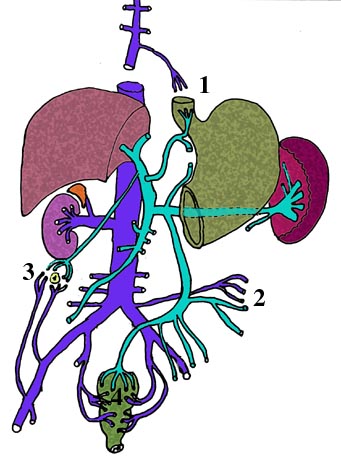
There are 4 points of anastomosis between the portal system and the Venal caval system and these areas are simply known as sites of portocaval anastomosis. They are:
1. Paraumbilical veins - superficial and inferior epigastric veins
2. Superior rectal veins - inferior and middle rectal veins
3. Colic twigs - lumbar segmental and renal venous branches
4. Esophageal veins of abdomen - esophageal veins of azygos system.
In the diagram below identify the 4 sites of portocaval anastomoses and label the parts of the portal system and the caval system.
|
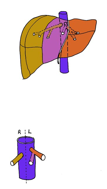
The liver can be divided in 3 different ways
Anatomically there are 4 lobes. However, there are 8 segments that can be identified based on their supply of unfiltered portal blood and hepatic arterial supply and bile salts production. Once filtered, these 8 segments drain into 3 main hepatic veins the Right one drains into the right half of the IVC and the middle and left ones drain into the left half of the IVC. Then blood is returned to the right atrium. |
|