CARDIOLOGY
DANIL
HAMMOUDI.MD
- CVD signs: Enlarged heart, swelling of the ankles or legs, unusual/ excess
weight gain, wounds that do not heal well.
- CVD symptoms: Chest discomfort (pain, pressure, squeezing,
heaviness, etc.) especially if brought on by exertion and relieved by
rest, shortness of breath with minimal exertion or upon lying down,
palpitation or irregular heart beats, severe dizziness or loss of
consciousness, sudden weakness or paralysis of one part of the body,
sudden slurring of speech or loss of vision, frequent nocturnal
urination, unusual and progressive fatigue, leg pain/ discomfort with
walking
Tips for clinical examination
§
Always
introduce yourself and be courteous to the patients
§
Ask for
permission before examination
§
Avoid hurting
the patient
§
Listen to the
instruction carefully
§
Do not forget
to observe the patient first for examples hearing aid or dermatitis etc.
§
Learn to
describe the physical findings before giving the likely diagnosis
§
Do not rush
in the examination. Remember
more than one signs may be present. (This may be associated or coincidental)
§
Do not panic
if you do not know the diagnosis. Give the physical findings and provide a list
of differential diagnosis (you may want to classify the pathology as congenital
or acquired; congenital may be inherited or non-inherited and acquired may be
traumatic, nepotistic, inflammatory, iatrogenic etc.)
§
Present your
findings confidently and look the examiner in the eyes. Avoid using words
"may be, I think, could be"
§
Treat each
case as new and do not let a bad case affect your subsequent examination
§
Avoid
derogatory such as syphilis, cancer or multiple sclerosis. Use euphemism or
medical jargons instead such as St.Louise's disease, neoplasm or demyelinating
diseases
§
Thank the
patients and examiners at the end of the examination even if the whole thing go badly.
Most cases
have vascular diseases involving the eye or the visual pathway. Therefore, look
for abnormal rhythm and left sided valvular disorders. The most likely cases
are:
- atrial
fibrillation
-
mitral stenosis
-
mitral incompetence
-
aortic stenosis
-
aortic incompetence
-
a mixture of the above
-
mechanical heart valve
-
Marfan's syndrome with aortic incompetence
-
carotid stenosis
- Vascular Exam (Arterial Bruits or
diminished pulses)
- Abdominal aorta
- Iliac artery
- Femoral artery
- Carotid artery
- Subclavian artery
- Local Signs of Peripheral Vascular Disease
- Decreased skin temperature
- Shiny skin
- Skin hairless over lower extremity (e.g.
shin)
- Dystrophic Toenails
- Distal extremity color change with
position
- Skin pallor
when leg elevated
- Skin rubor
when leg dependent
MURMURS
BASAL SYSTOLIC MURMUR:
- VALVULAR AORTIC STENOSIS
- HYPERTROPHIC SIBAORTIC STENOSIS
- SUPRAVALVULAR AORTIC STENOSIS
- HYPERTENTION OR ATHEROSCLEROSIS
- VALVULAR PULMONIC STENOSIS
- INFUNDIBULAR PULMONIC STENOSIS
- ATRIAL SEPTAL DEFECT AT OSTRIUM SECUMDUM
- COARTATION OF THE AORTA
- CARDIOPULMONARY M
- BENIGN MURMUR
MIDPRECORDIAL MURMUR:
- VENTRICULAR SEPTAL DEFECT
- TRICUSPID REGURGITATION
APICAL SYSTOLIC MURMUR
- MITRAL REGURGITATION
- PAPILLARY MUSCLE DYSFUNCTION
- BENIGN MURMUR
SYSTOLIC CLICK
- AORTIC EJECTION CLICK [
1. ANEURYSM IN ASCENDING AORTA,
2. COARTATION OF AORTA,
3. HTA WITH
AORTIC DILATION,
4. VALVULAR AORTIC STENOSIS,
5. AORTIC
REGURGITATION
- PULMONIC EJECTION CLICK
·
PULMONARY VALVE STENOSIS
·
PULMONARY ARTERY
DILATION
·
[DECREASE OR
DISAPPEARS WITH INSPIRATION
·
INCREASED WITH
EXPIRATION
APICAL SYSTOLIC MURMUR
WITH MIDSYSTOLIC CLICK:
·
MITRAL LEAFLET
PROLAPSE
Areas of the heart
Mitral
- apex
- should be 5th intercostal space and in mid-clavicular line
Tricuspid
- lower left sternal edge
Pulmonary
- upper left sternal edge ie left of manubrium
Aortic
- upper right sternal edge ie right of manubrium
Heart Sounds
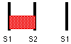
1st Heart Sound, S1
This signals the onset of
systole and is caused by the closure of the mitral and tricuspid valves with
concomitant tensing of the left ventricular wall. The mitral valve closes
slightly before the tricuspid valve but the two sounds are usually merged.
Splitting is marked in tricuspid stenosis or right bundle branch block. NB The
1st sound can be identified by palpating the carotid pulse while auscultating -
the upstroke of the carotid pulse closely follows the 1st heart beat.
- S1 is loud in
mitral stenosis -> palpable tapping apex beat; also in
tachycardia or hyperdynamic circulation
- S1 is soft in
mitral regurgitation (and also when the PR interval is long);
also in bradycardia and LVF
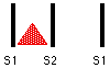
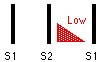
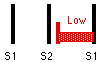
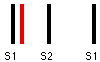
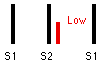
2nd Heart Sound, S2
This separates systole and
diastole. The sound is made by the closure of aortic and pulmonary valves. The
aortic valve closes before the pulmonary valve and this splitting of the second sound is heard particularly
during inspiration, as more blood is drawn into the right
ventricle, a normal phenomenon. (This occurs because right heart venous return
varies with respiration). During expiration the split sounds of S2
resynchronise. The sound of pulmonary
valve closure (P2) is best heard over the pulmonary area
as it is much quieter than that of aortic valve closure (A2).
- Wide splitting occurs in
- right bundle branch block
- pulmonary stenosis
- Wide and fixed splitting (ie not varying with respiration) occurs when there is an atrial septal defect
- Reversed splitting (ie
splitting increasing on expiration) occurs in
- systemic hypertension
- left bundle branch block
- aortic stenosis
- NB In aortic
stenosis A2 is often soft or absent -> "single
component 2nd sound"
- A2 is loud in systemic hypertension
- P2 is loud in pulmonary hypertension
- P2 is soft in pulmonary stenosis
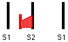
3rd & 4th Heart Sounds
These are low pitched (so
heard best with the bell) and not usually heard.
If
either S3 or S4 is very loud a gallop/triple rhythm will
result.
Loud
S3 rhythm = S1----S2--S3 ie
crotchet-quaver-quaver
Loud
S4 rhythm = S4-S1-----S2 ie
semiquaver-crotchet-crotchet
3rd Heart Sound, S3
This is produced by rapid
ventricular filling and occurs in early-mid diastole ie soon after S2
Occurs
normally in young fit adults with bradycardia eg athletes
Occurs
abnormally in
- patients with heart failure
- left heart failure - S3 heard best
in mitral area
- right heart failure - S3 heard best
in tricuspid area
- patients with high ventricular
filling pressure
- mitral regurgitation
- constrictive pericarditis
- higher pitched and in early diastole =>
"pericardial knock"
- occurs because ventricular filling is suddenly
curtailed by constriction of pericardium
4th Heart Sound, S4
This is an atrial sound, occurring just before S1.
It is always abnormal as it
represents atrial contraction against a stiffened ventricle eg due to aortic stenosis or hypertensive heart disease. It may also
occur in heart failure.
Added Sounds
Ejection systolic
clicks - usually due to aortic stenosis and systemic hypertension
but can be to pulmonary stenosis or
pulmonary hypertension.
Mid-systolic
clicks occur in mitral valve prolapse.
Opening
snap may occur in mitral or tricuspid stenosis.
Prosthetic
valves make noises on opening and closing.
A pericardial friction rub is a creaking
sound heard in systole or diastole which suggests pericarditis (could be viral,
post MI, due to CTdisease, trauma or uraemia) so look for an associated
pericardial effusion.
Murmurs
Murmurs are caused by
turbulent blood flow; this may be due to a stenotic or regurgitant valve
producing a high velocity jet; alternatively it may be due to increased flow
velocity in a normal vessel or to normal velocity flow in a dilated or
distorted vessel; non-pathological causes include the murmur of the
hyperdynamic system in pregnancy or a minor anatomical distortion with no
pathological consequences (innocent murmur).
Bell
is good when listening to low-pitched sounds eg mitral stenosis. Diaphragm is
good when listening to high-pitched sounds eg aortic regurgitation.
How to amplify murmurs
Left
heart murmurs are accentuated in expiration; right
heart murmurs are accentuated in inspiration
Performing
the Valsalva manoeuvre (get patient to strain silently)
- amplifies the murmurs of mitral valve prolapse and hypertrophic
obstructive cardiomyopathy
- softens the murmurs of mitral regurgitation and aortic stenosis
NB Sudden squatting has the
opposite set of effects to performing the Valsalva manoeuvre.
Mitral
stenosis will be heard better if the patient rolls into the left lateral
position.
Aortic
regurgitation will be heard better if the patient leans forward.
Murmurs
are also amplified by exercise due to increase in cardiac output.
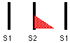
Systolic murmurs
1) Ejection systolic murmur = crescendo-decrescendo
murmur
This
originates from the outflow tract, waxing and waning with changing
intra-ventricular pressure.
It
may be a flow murmur, common in childhood and pregnancy, or a pathological
murmur as in
- aortic stenosis
- aortic sclerosis
- hypertrophic obstructive cardiomyopathy
- pulmonary stenosis
- atrial septal defect
2) Pansystolic murmur
Of
uniform intensity and merges with S2 although it may obscure both S1
and S2. It is usually caused by jets passing from a high pressure
chamber to a low pressure chamber during systole ie mitral or tricuspid regurgitation. A pansystolic murmur also occurs
when there is a ventricular septal defect.
3) Late systolic murmur
This
is caused by mitral valve prolapse or
papillary muscle dysfunction. It is a high-pitched murmur of even
intensity which starts halfway through systole with a mid-systolic click and
terminates with A2. NB If the papillary muscle rupture has occurred
posteriorly then this murmur will be loudest in the aortic area (instead of the
mitral area where it is heard most of the time).
Diastolic murmurs
1) Early diastolic murmur
This
is high-pitched and usually only heard as the "absence of silence" in early
diastole.
It
occurs due to aortic or pulmonary
regurgitation. The aortic regurgitation murmur is usually soft and
is best heard with the patient leaning forward and in expiration.
NB When pulmonary regurgitation is due to pulmonary
hypertension caused by mitral stenosis then the early diastolic murmur is
called a Graham-Steel murmur.
2) Mid-diastolic murmur
This
is low-pitched and rumbling; it starts after an opening snap.
Caused
by
- mitral stenosis (common)
- exhibits pre-systolic accentuation in sinus
rhythm
- amplified by rolling the patient into the left
lateral position
- rheumatic fever
- thickens mitral valve leaflets
- = Carey
Coombs' murmur
- aortic regurgitation
- regurgitant jet causes fluttering of anterior
mitral valve cusp
- = Austin
Flint murmur
- tricuspid stenosis (rare)
- large atrioseptal defect
Other murmurs
1) Continuous, machinery murmur of patent ductus
arteriosus.
2) Musical or "mewing" murmurs -
characteristic of a hole in an aortic
valve cusp due to endocarditis
3) Innocent murmur
- common in children or in pregnancy
- loudest in pulmonary area
- often low pitched and low intensity
- check them out with Echocardiograms
and Doppler studies
- ? Marfan's -> aortic regurgitation
- ? Rheumatological disorders eg ank. spond -> aortic
regurgitation
- ? Down's -> ASD or VSD
- ? Turner's -> coarctation of aorta
- ? Thyrotoxic -> predisposed to AF and high output heart failure
- ? Alcoholism -> dilated cardiomyopathy
Hands
- Clubbing
- Cyanotic congential heart disease
- Infective endocarditis
- Peripheral cyanosis
and Perfusion
- Endocarditis is suggested by
- Splinter haemorrhages
- Osler's nodes = tender lumps in pulp of fingertips
- Janeway lesions = red macules on wrist and hand
- Nicotine stains - peripheral vascular disease
Radial pulse - for rate and rhythm
- Irregularly irregular = atrial fibrillation (or multiple ectopics)
- Regularly irregular = 2nd degree heart block
- Water hammer pulse (= Collapsing)
- strong radial pulse that taps hand on lifting
of arm
- indicates wide pulse pressure of aortic
regurgitation
- Bounding pulse
- CO2 retention
- Liver failure
- Sepsis
- Small volume thready pulse = shock
- Radio-radial delay -
suggests coarctation or dissection
- Arterio-venous fistulae - buzzing - for dialysis
Carotid pulse - for character
- Normal
- Small volume - in low
output states eg heart failure, shock, mitral stenosis
- Small Volume And Slow Rising pulse = aortic stenosis
- Collapsing (rapid up
and rapid down) in aortic regurgitation (also AV fistula or hyperdynamic
cicrulation)
- Bisferiens =
collapsing and slow rising occurring in mixed aortic disease
- Pulsus alternans - LVF
- Jerky - hypertrophic
cardiomyopathy
- Pulsus Paradoxus -
pulse weakens in inspiration - indicates tamponade or constrictive
pericarditis
Face
- Malar flush = mitral stenosis (also present in mixed mitral disease)
- ? Jaundice - poss. prosthetic valve causing mild haemolysis
Eyes
- Argyll-Robertson pupil
- pupil constricted, does not react to light but
does to accomodation
- "the prostitute's pupil accomodates but
does not react"
- think of syphilitic aortic regurgitation &
poss. aneurysm
- Xanthelasmata or corneal arcus = hyperlipidaemia
Mouth
- Cyanosis
- High arched palate of Marfan's -> aortic regurgitation
- Mucosal petechiae -> infective endocarditis
JVP
Inspection of precordium
- Scars
- Deformity
- Pulsation
- Pacemaker boxes
Palpation
- Thrills
- Heaves
- parasternal heave of RVH
- apex beat may be
- tapping (quick and light) - mitral stenosis
- thrusting (diffuse and long) - mitral regurgitation
- heaving (sharp and firm) - LVH & aortic stenosis
- Apex beat should be 5th intercostal space mid-clavicular line
Auscultation
- Remember to roll into left lateral position and to sit forwards
- Remember to listen on inspiration and on held expiration
Chest
- Listen at lung bases for fine
inspiratory creps of pulmonary oedema (LVF)
Sacral oedema
Abdomen
- Hepatomegaly - RVF
- Pulsatile hepatomegaly - tricuspid regurgitation
- Splenomegaly - endocarditis
- Pulsatile mass (not liver) - abdominal aneurysm
- Femoral arteries, radio-femoral delay (coarctation of aorta) and
femoral bruits
- Also can listen for renal bruits
Peripherally
- Peripheral pulses
- Pitting oedema
- Peripheral vascular disease - cold feet, gangrene
- Varicose veins
Finally
- BLOOD PRESSURE
- narrow pulse pressure indicates aortic stenosis
- wide pulse pressure indicates aortic
regurgitation
- drop of > 10mm Hg in inspiration indicates
pulsus paradoxus and either
tamponade or constrictive pericarditis
- Fundi
- hypertensive change
- grade I copper wiring of arteries
- grade II arteriovenous nipping
- grade III flame or blot haemorrhages, cotton
wool exudates
- grade IV papilloedema
- Roth' spots = retinal vasculitis indicative of
endocarditis
- Urine - haematuria may indicate endocarditis
- Temperature chart - endocarditis
The Signs of Different Conditions
Aortic Stenosis (Uncomplicated)
- Observation - more likely to be male
- Hands - nil
- Radial pulse
- normal
- AF
(irregularly irregular)
- Carotid pulse - slow
rising pulse
- Face - nil
- JVP - normal
- Thrills
- aortic areas
- 2nd R intercostal space (classical aortic
area)
- 4th L intercostal space/sternal edge whch is
along the line of LV ejection
- over apex (along line of LV ejection)
- Apex
- normal position 5th intercostal space in
mid-clavicular line
- heaving character due to LVH
- Auscultation
- 1st HS normal
- Ejection systolic click may precede murmur
- Ejection systolic murmur
- loudest sitting forward
- loudest in expiration
- radiates to carotids and apex
- 2nd HS
- may be soft
or absent
- paradoxical splitting may occur during
expiration such that P2 occurs before A2
but A2 may not be audible anyway
- No diastolic murmurs
- 4th HS may be present just before 1st HS
- Peripherally nil of note
- Blood pressure
- narrow pulse pressure eg 120/80 or 110/80
Mitral Regurgitation (Uncomplicated)
- Observation - nil
- Hands
- look for signs
of endocarditis ie splinter haemorrhages (transilluminate
them), Osler's nodes, Janeway lesions
- Radial pulse - may find AF
- Face - normal in uncomplicated mitral regurgitation (but malar flush of mitral stenosis if mixed mitral
disease)
- Carotid
- usually normal character
- in severe disease may get a small volume jerky pulse due to
shortened ejection volume and time
- JVP
- usually normal
- raised if subsequent pulmonary hypertension
- Thrill possible in mitral area
- Heaves
- in severe regurgitation may develop a parasternal heave due to left atrium
enlargement
- may also have RV Heave if pulmonary hypertension has developed
- Apex
- displaced laterally and down
- thrusting
- Auscultation
- 1st HS - soft or normal
- Pan-systolic murmur
- loudest at apex in left lateral position
- radiates to the axilla
- may obscure aortic component of 2nd HS
- 2nd HS may be obscured (but if there is pulmonary hypertension it could be loud and
late)
- 3rd HS often present
Abnormalities of the JVP
1) Raised JVP with normal waveform
- right heart failure
- fluid overload
- bradycardia
2) Raised JVP with absent pulsation
- SVC obstruction - full dilated jugular veins, no pulsation,
oedematous face and neck
3) Large a wave
- tricuspid stenosis - atria contracts against stiff tricuspid and so
pressure in atria rises higher than normal
- pulmonary hypertension - there are generally higher pressures on
the right side of the heart
- pulmonary stenosis
4) Extra-large a wave = Cannon wave
Occurs
when atrium contracts against closed tricuspid eg
- complete heart block
- atrial flutter
- single chamber pacing
- nodal rhythm (AV node is in charge)
- ventricular extra-systole
- ventricular tachycardia
ie any condition in which
the atria and the ventricles are not conducting in appropriate rhythm
5) Absent a wave
- atrial fibrillation
6) Systolic waves = combined c-v waves = big v waves
- tricuspid regurgitation (c-v wave because the pressure in the right
atrium is raised throughout ventricular systole - tip is to watch for
earlobe movement!)
7) The slow y descent occurs in tricuspid
stenosis (if the HR is so low as to allow the length of descent to be
appreciated!)
8) Paradoxical JVP = Kussmaul's sign
Normally
the JVP should rise on expiration and fall on inspiration.
When
the JVP rises on inspiration it indicates
- pericardial effusion
- constrictive pericarditis
- pericardial tamponade
Right Ventricular Failure
·
R side heart
failing
·
Pooling blood
IVC< V
·
Raised JVP
·
Hepatomegaly
·
Ankle oedema
·
Cyanosis
·
Signs of what
caused it
Pericardial Disease
·
Pericarditis
o
friction rub
·
Tamponade
o
JVP raised
o
very reduced
heart sounds
§
apex beat
impalpable
o
hypotension
Aortic Stenosis
·
Narrowing
imposes a pressure load on LV
·
Restricts LV
outflow
·
Midsystolic
harsh ejection murmur
·
Max at aortic
area
·
Loudest sitting
and expiration
|
Blood Pressure Classification in Adults |
||
|
Category |
Systolic |
Diastolic |
|
Normal |
<130 |
<85 |
|
High Normal |
130-139 |
85-89 |
|
Mild Hypertension |
140-159 |
90-99 |
|
Moderate Hypertension |
160-179 |
100-109 |
|
Severe Hypertension |
180-209 |
110-119 |
|
Crisis Hypertension |
>210 |
>120 |
What does a blood pressure of 170/80 indicate?
Pure Systolic Hypertension
Have the patient roll on their left side.- Listen with the bell at the apex.
- This position brings out S3
and mitral murmurs.
- Have the patient sit up, lean forward, and hold
their breath in exhalation.
- Listen with the diaphragm at
the left 3rd and 4th interspace near the sternum.
- This position brings out
aortic murmurs
|
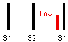
Murmurs and Extra Sounds |
||
|
|
|
Systolic Click |
|
|
|
Opening Snap |
|
|
|
|
|
Murmur Grades |
||
|
Grade |
Volume |
Thrill |
|
1/6 |
very faint, only heard
with optimal conditions |
no |
|
2/6 |
loud enough to be obvious |
no |
|
3/6 |
louder than grade 2 |
no |
|
4/6 |
louder than grade 3 |
yes |
|
5/6 |
heard with the stethoscope
partially off the chest |
yes |
|
6/6 |
heard with the stethoscope
completely off the chest |
yes |
- Occlusion Location
- Aortoilliac Occlusive Disease (Leriche's
Syndrome)
- Bilateral leg diminished pulses throughout
- Slow wound healing legs
- Impotence
- Iliofemoral Occlusive Disease
- Unilateral leg diminished pulses throughout
- Buttock claudication may be present
- Femoropopliteal Occlusive Disease
- Thigh and calf claudication
- Normal femoral pulses in groin
- Ankle-Brachial ratio > 0.9: Normal
- Ankle-Brachial ratio 0.5 to 0.9:
Claudication
- Ankle-Brachial ratio < 0.5: Resting
ischemic pain
Chronic Venous
Insufficiency
|
|
Risk Factors
- Obesity
- Congestive Heart Failure
- Diabetes Mellitus
- Symptoms and Signs
- Initial Changes
- Varicose veins
- Tan or reddish brown skin color changes
- Weeping and excoriated skin
- Pedal edema
- Later Changes
- Lipodermatosclerosis
- Induration at
medial ankle to mid-leg
- Advanced Changes
- Brawny edema above and below fibrotic area
- Ulcerations
- Complications
- Venous Stasis Ulcers
- More common in older women
- Chronic and often recurrent
- Postphlebitic Syndrome
- Chronic leg edema
- Deep Venous Thrombosis
- Pigmentation
- Ulceration
- Diagnosis
Abdominal Aortic Aneurysm
Causes
- Associated with Atherosclerosis in only 25%
of patients
- Aortic Dissection
- Mycotic Infection
- Cystic Medial Necrosis
- Ehlers-Danlos Syndrome
- Asymptomatic in 75% of AAA
- Abrupt onset severe pain unrelieved by
position change
- Suggestive of aneurysm enlargement or
rupture
- Aortic Rupture (20% present ruptured)
- Free Intraperitoneal Rupture
(Catastrophic)
- Acute pain
- Cardiovascular
Collapse
- Sudden Death
- Sentinal Bleed (small posterolateral wall
tear)
- Acute pain
(constant)
- Syncope
- Pulsatile abdominal mass
- Hemodynamically
stable with tachycardia
- Needs
Emergent Intervention before full rupture
Findings suggestive of occlusion
- Proximal lower extremity pressures less
than arms
- Proximal femoral pressure <20 mmHg over
brachial
- Drop in doppler pressures between segments
>20-30 mmHg
Edema evaluation
|
|
- Examiner impresses thumb into skin over
bony surface
- Tibia
- Fibula
- Sacrum
- Withdraw thumb
- Measure depth of pit and record in
millimeters
- Interpretation
- Pitting Edema
- Non-Pitting edema (Brawny edema)
- Myxedema
- Chronic inflammation
- Chronic Venous Stasis
- Edema Distribution
- Dependent Edema (fluid shift in response to
gravity)
- Standing patient accumulates fluid in feet
and ankles
- Bed-bound patient collects fluid
posteriorly (sacrum)
- Chronic Leg edema (Brawny edema)
- Tissue becomes fibrotic and fails to pit
Chest Pain
- A heart attack.
- Lung problems like pneumonia, bronchitis, or an
injury.
- A hiatal hernia - known in medical terms as
gastroesophageal reflux disease (GERD).
- Heartburn.
- Shingles.
- A pulled muscle.
- Mitral valve prolapse. A common disorder, especially
in women, in which the mitral valve of the heart fails to close properly.
In most people, this is not a serious problem.
- Anxiety.
- Swallowing too much air.