USMLE STEP 1 REVIEW

DANIL HAMMOUDI.MD

SINOE MEDICAL ASSOCIATION


biochem

Alkaptonuria and Ochronosis

Alkaptonuria is a rare disease in which the body does not have enough of an enzyme called homogentisic acid oxidase (HGAO). It is a genetic disease, meaning that it is inherited from a family member.

Because normal amounts of the HGAO enzyme are missing, homogentisic acid (HGA) is not used and builds up in the body. Some is eliminated in the urine, and the rest is deposited in body tissues where it is toxic. The result is ochronosis, a blue-black discoloration of connective tissue including bone, cartilage, and skin caused by deposits of ochre-colored pigment.

Patients with alkaptonuria are usually not aware of the disease until about age 40 when symptoms are present. Dark staining of the diapers sometimes can indicate the disease in infants, but usually no symptoms are present until much later in life.

Alkaptonuria and ochronosis affect many body systems, as described below.

  • Skeletal (bones and cartilage)--The knees, shoulders, and hips are most affected; arthropathy (diseased joints) is common. Deposits of pigment cause cartilage to become brittle and eventually to fragment (break apart).
  • Cardiovascular (heart and blood vessels)--The aortic and mitral heart valves are most affected. Ochronotic granules can cause valves to calcify or harden. Pigment deposits also can lead to the formation of atherosclerotic plaques (hard spots in arteries) containing cholesterol and fat.
  • Genitourinary (genital and urinary systems and organs)--In men, the prostate is most commonly affected. Pigment deposits can form stones in the prostate.
  • Respiratory (organs and structures involved in breathing)--Heavy pigment deposits in the cartilage of the larynx (voice box), the trachea (windpipe), and the bronchi (air passages to the lungs) are common.
  • Ocular (eyes)--Vision is not usually affected, but pigmentation in the white part of the eye is evident in most patients by their early forties.
  • Cutaneous (skin)--Effects are most noticeable in areas where the body is exposed to the sun and where sweat glands are located. Skin takes on a blue-black speckled discoloration. Sweat can actually stain clothes brown.
  • Other--The teeth, central nervous system (brain and spinal cord), and endocrine organs (which make hormones) also may be affected.

Arthropathy (joint disease characterized by swelling and enlarged bones) and discoloration of the skin cause the greatest disability.

Usually a physician can diagnose alkaptonuria based on symptoms of joint discomfort and skin discoloration. The diagnosis is confirmed by verifying family history of the disease, examining skin cells, and testing the urine. Urine left standing for several hours will turn brownish black if a patient has alkaptonuria.

Diets low in protein--especially in amino acids, phenylalanine (found in aspartame), and tyrosine--help reduce the levels of HGA, thereby lessening the amount of pigment deposited in body tissues. Symptoms of alkaptonuria (e.g., arthropathy, cardiovascular disease) are treated when possible. Unfortunately, the course of the disease remains unchanged, and no cure is available. However, patients tend to have a normal life span and die of causes comparable to those of the general population.

Alkaptonuric ochronosis represents an autosomal recessively transmitted inborn error of metabolism. It is the result of a deficiency of the enzyme homogentisate 1,2-dioxygenase1 caused by mutations in the homogentisate 1,2-dioxygenase gene located to a 16-cM region of the 3q2 chromosome.2 The resulting elevation of serum homogentisic acid levels causes massive excretion of homogentisic acid in the urine, which turns black on oxidation.3 Homogentisic acid deposition in connective tissue throughout the body causes the tissue to become pigmented, a feature termed ochronosis by Virchow in 1866.4 Deposition in the intervertebral disks and joint cartilage results in ochronotic spondylopathy and arthropathy.3 In the eye, ochronotic pigmentation, histopathologically seen as amorphous and curlicue deposits, occurs primarily in the conjunctiva and sclera in the interpalpebral fissure.5 To our knowledge, the literature does not mention any effects on visual function or corneal topography. In the case presented here, the enlarging perilimbal ochronotic scleral lesions nasally and temporally were associated with progressive peripheral corneal thinning and astigmatism in the axis of the lesions.


aka: alkaptonuria

  • autosomal recessive
  • excessive homogentisic acid
    • no homogentisic acid oxidase
    • part of phenylalanine / tyrosine pathway
  • alkaptonuria: brown/black urine
  • ochronosis: brown/black pigmentation of skin, mucous membranes
  • pigment deposition in articular cartilage of joints
    • ==> chronic arthritis (usually evident in 4th decade)
  • narrowed, calcified intervertebral disks
  • renal calculi
  • nephrocalcinosis

Ochronosis is one of common side effect of long term high potency hydroquinone treatment . Clinically there is slate gray to browish discoloration of the face. Under microscope examination there is homogenous brownish material in the dermis. This may be denatured collagen or other proteins. This condition is
rather difficult to treat with medication. The best treatment is now by doing laser ablation with either Carbondioxide laser or Erbium :YAG laser. Laser will remove skin down to the dermis follow by regeneration of new healthy skin. There usually is post treatment hyperpigmentation which will eventually fade away. This condition is one of the strong reason why we should not use hydroquinone for long term treatment of melasma


It is a hereditary enzyme deficiency of the homogentisic acid oxidase in the liver and the kidneys. The gene is transmitted in a recessive autosomal mode. The incidence of alkaptonuria is 1 per million with the highest prevalence in Slovakia by inbreeding (10). Homogentisic acid (HGA) is an intermediary metabolite of the aromatic acids phenylalanine and tyrosine (Figure 1).

Figure 1 : Biochemical disorder of alkaptonuria.

The ochronotic pigment granules have a high affinity for fibrillary collagens which are surrounded by abundant mucopolysaccharide ground substance. The most outstanding example are the hyaline cartilages of the joints. HGA is excreted in the urine (4-8 g/day). Upon oxidation of HGA the excreted urine is discolored black (3). Ochronotic pigment accumulation in the cartilage of the external ear.

It was Scribonius who described the first known case of ochronosis in 1584. He mentioned a schoolboy which passed urine black as ink (3). In 1902, Albrecht and Zdareck discovered the link with alkaptonuria.

 
 Radiograph of the spine and pelvis demonstrating the waferlike calcifications of the intervertebral disks and ankylosing of the spine and sacroiliac joints (open arrows). Advanced degenerative arthritis is seen in the hips (closed arrow).

Ochronotic Pigment Deposits in the Sclerae.


Discussion

The clinical manifestations are caused by the accumulation of HGA in the tissues rich of fibrillary collagen. The binding by polymerisation and auto-oxidation of HGA is irreversible(16). Ochronotic arthropathy is caused by a deposition of the ochronotic pigment in the articular cartilage. This leads to a brittleness and fragmentation of the cartilage causing a non-specific synovitis. Also typical is the ochronotic spondylolysis in the lumbar spine. The stiffness of the back slowly progresses to a complete rigidity with flattening of the lumbar lordosis and dorsal hyperkyphosis resembling ankylosing spondylitis. In contrast to ankylosing spondylitis, syndesmophytes or an annular ossification with a bamboo pattern do not occur. There is a progressive desintegration and ossification of the nucleus pulposus, causing a diminished length sometimes more than 15 centimetres. There can also be a restriction of breathing because of affected costovertebral joints. Pain, stiffness, crepitation, flexion contractures and limitation of motion are the most common clinical features of the periferal ochronotic arthropathy. Deposition of calciumpyrophosphate cristals with attacks of pseudo-gout are associated with ochronotic arthropathy. The radiographic findings are not characteristic : a non-specific osteo-arthritis, chondrocalcinosis, peri-articular calcifications and osteochondromathosis.

The most important clinical signs of ochronosis are the pigmentations of the sclerae, auricles and the interphalyngeal and metatarsal joints. The otoscopic examination also demonstrates black cerumen. The earbones and the membranum tympanum have deposits of ochronotic pigments causing tinitus and diminished hearing.

The cardiovascular examination reveals an aortic murmur as a result of aortic stenosis and calcification. The deposits of ochronotic pigments in the valves could be an occasion for dystrophic calcification. Genito-urinary tract obstruction occurs in 60 % of ochronotic patients. Rectal examination reveals a nodular prostate in consequence of prostatic calcifications.

The corner stones of diagnosis are the examination of the urine (black color) and the histopathological examination with hematoxylin-eosin staining confirming the ochronotic pigments in the tissues.

There is no cure for ochronotic arthropathy. The enzyme deficiency is not to be treated yet (gentherapy in the future ?). Some physicians prescribe high doses of ascorbic acid to prevent the interaction of the ochronotic pigment with the tissues.(5) Unfortunately, the progress of the disease is not interrupted by this treatment. There only exists a symptomatic treatment : analgetics, NSAID, physical therapy, orthopaedic supports and intra-articular corticoid infiltration, especially in the knee. Finally, arthroplasty of the hip and knee is often necessary.


anatomy


if the urethra were damaged in the bilb of the penis , urine could extravase into : superficial perineal space


the superficial perineal space and its contents.


Definition: a potential space between the membranous layer of the subcutaneous tissue

and perineal membrane (= inferior fascia of the UG diaphragm)


Relationships:

·        Superiorly: inferior fascia of the urogenital diaphragm

·        Inferiorly: superficial perineal fascia (= continuation of superficial fascia of the abdomen)


Contents:

·        Right and left corpus cavernosum (of penis/clitoris)

·        Crus of each corpus cavernosum (of penis/clitoris)

·        Bulb of penis/vestibule

·        Superficial transverse perineal muscles

·        Bulbospongiosus muscle

·        Male: overlying the corpus spongiosum

·        Female: beneath the labia majora

·        Ischiocavernosus muscle

·        Male: overlying the corpus cavernosum

·        Female: beneath the labia majora

·        Male only:

·        Corpus spongiosum

·        Penile urethra

·        Female only:

·        Greater vestibular glands


Blood vessels:

·        Branches of the internal pudendal vessels (deep and dorsal arteries of penis/clitoris)


Lymphatics: superficial inguinal lymph nodes


Innervation:

·        Branches of the pudendal nerves (dorsal nerve of penis/clitoris, posterior scrotal/labial nerves)


Male Urogenital Triangle

Make a midline incision beginning at the shaft or posterior portion of the penis and split the scrotum into right and left halves. Identify the superficial and posterior scrotal branches of the perineal nerve (terminal branches of the pudendal nerve) and the perineal and posterior scrotal vessels (branches from the internal pudendal vessels). Continue to reflect the skin flaps laterally. You are now in the superficial perineal space/pouch.

The contents of the superficial perineal pouch include three paired muscles and portions of the penis. Identify the ischiocavernosus muscle covering the crura of the penis, an extension of the corpus cavernosus originating from the ischial tuberosity. Identify the bulbospongiosus muscle covering the bulb of the penis, the first part of the corpus spongiosum. This muscle arises from the median raphe and the perineal body. Identify (if you can) the superficial transverse perineal muscle. This muscle runs from the ischial tuberosity to the perineal body. It is usually very small, so try looking for any fibers coursing transversely across the posterior boundary of the perineal membrane.

Between the crus and the bulb of the penis you can see the perineal membrane, the fascia of the UG diaphragm. Superior to this fascia are the deep transverse perineal muscle and the sphincter urethrae (external urinary sphincter), the muscles of the UG diaphragm. In the male the bulbourethral (Cowper’s) gland also lie in this space.

Q1. Ducts from Cowper’s gland open into the _________.

Remove the superficial fascia of the penis and observe the deep (Buck’s) fascia of the penis. Open the deep fascia on the dorsum of the shaft of the penis and identify the deep dorsal vein, the paired dorsal arteries, and the paired dorsal nerves.

Study the components of the shaft of the penis; the paired corpora cavernosa and the corpus spongiosum. Separate the erectile tissue and observe the glans penis (distal expansion of the corpus spongiosum) and the spongy or penile urethra. Make a cut longitudinally and observe the spongy urethra and its distal expansion, the navicular fossa.

Cut open the scrotum and expose the testis. Identify the epididymis and follow the course of the vas deferens from the scrotum to the superficial inguinal ring. Identify the tunica vaginalis covering the testis and epididymis. Open the testis and observe the thick tunica albuginea encasing the testis and the septa containing the seminiferous tubules. Try to locate the rete testis and efferent ducts. Distinguish the caput (head), corpus (body) and cauda (tail) portions of the epididymis. Finally, find the testicular artery and appreciate the venous (pampiniform) plexus.


Female Urogenital Triangle

Make an incision between the anus and the vagina, and then anteriorly around the right and left labia majora toward the pubic symphysis. Continue to reflect the skin flaps laterally. You are now in the superficial perineal space/pouch. Identify the superficial and posterior labial branches of the perineal nerve (terminal branches of the pudendal nerve) and the perineal and posterior labial vessels (branches from the internal pudendal vessels).

HINT: Identify the portions of the clitoris in the superficial pouch and look for the muscles associated with these tissues - the crura (alongside the ischialpubic ramus) and the bulb of the vestibule. (midline) (Grts. 3.59, 3.77; Net. PL. 356)

The contents of the superficial perineal pouch include the three-paired muscles, part of the clitoris, and the bulb of the vestibule. Identify the ischiocavernosus muscle covering the crura of the clitorus, an extension of the corpus cavernosus originating from the ischial tuberosity. Identify the bulbospongiosus muscle covering the bulb of the vestibule. This muscle arises from the median raphe and the perineal body. Attached posterior to the bulb of the vestibule are the greater vestibular (Bartholin’s) glands. (In most elderly, postmenopausal female cadavers, the accessory organs and glands are atrophic and not readily evident). Identify (if you can) the superficial transverse perineal muscle. This muscle runs from the ischial tuberosity to the perineal body. It is usually very small, so try looking for any fibers coursing transversely across the posterior boundary of the perineal membrane.

Between the crus and the bulb of the vestibule you can see the perineal membrane, the fascia of the UG diaphragm. Superior to this fascia are the deep transverse perineal muscle and the sphincter urethrae (external urinary sphincter), the muscles of the UG diaphragm. In the female, the the vagina passes through the UG diaphragm and the membranous urethra is situated anterior to the vagina.

Identify the deep dorsal vein, the paired dorsal arteries and the paired dorsal nerves on the dorsum of the clitoris. Study the components of the erectile tissue of the clitoris, which consist of only two erectile bodies, the paired corpora cavernosa. The corpora cavernosa begin as the crura of the clitorus attached to the ischiopubic rami and end in the midline as the body of the clitoris and the glans.

ANS. 1. spongy (penile) urethra


urine could extravase into the anterior thigh if the urethral ruptured occurred in the superficial peritoneal pouch.


Into the peritoneal cavity =č ureter damage


Ischioanal fossa or pudendal canal =č urethral rupture


THE PELVIS

The "pelvis" itself is where the trunk and lower limbs meet and is divided into a MAJOR or FALSE pelvis and a MINOR or TRUE pelvis.

  • The PELVIC BRIM is the same as the SUPERIOR PELVIC APERTURE and the PELVIC INLET. It is the region bounded by the arcuate line and separates the true or minor pelvis from the false or major pelvis.

- The false pelvis contains structures that are part of the abdomen.

BOUNDARIES OF THE TRUE PELVIS

  • Lateral wall: obturator internus muscle
  • Posterior wall: sacrum, parts of the ilium, sacroiliac joint, piriformis muscle
  • Anterior wall: pubic bones and parts of obturator internus muscle
  • Floor: pelvic diaphragm

PELVIC DIAPHRAGM- the "funnel" Dr. Tucker placed in the hip bones.

Composed of:

(3) Levator ani muscles: puborectalis, pubococcygeus, and iliococcygeus

Coccygeus muscle

Its function include: supporting the abdominal and pelvic viscera, assisting the abdominal muscles in increasing abdominal pressure (for pushing hard during childbirth or even when on the toilet). The puborectalis forms the puborectal sling which is part of the external anal sphincter.

SACRAL PLEXUS = L4-S4

  • - is found on the anterior surface of the piriformis muscle
  • - part of L4 and L5 make up the Lumbosacral trunk
  • - major branches include: sciatic n., pudendal n., and superior and inferior gluteal n.
  • - the only new one is the PUDENDAL n. which exits the greater sciatic foramen, loops around the sacrospinous ligament, and enters the lesser sciatic foramen.
  • - Pudendal n. innervates the anal canal muscles and external genitalia or the perineum.

UROGENITAL DIAPHRAGM = thin sheet of muscle that spans the pubic arch

- made of deep transverse perineal muscle and voluntary sphincter urethra muscle (you should know what this does)

-covering the UG diaphragm is the perineal membrane (deep fascia) 

PELVIS BLOOD SUPPLY

URINARY BLADDER

  • -Urine enters the bladder from the two ureters that come from the kidney and enter the bladder at an oblique angle. Why is this angle important?
  • -Vesico- is the prefix meaning bladder.
  • - Make sure you know the parts of the bladder in your syllabus.
  • - The trigone is the smooth part of the bladder that is non-stretchable.
  • - The urethra is different in males and females.

In females, it is short and ends at external urethral orifice in the vestibule of the vagina.

In males, it is much longer and divided into 3 parts: membranous, prostatic and spongy.

INTERNAL GENITALIA

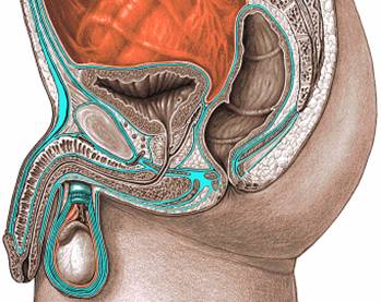
MALE = DUCTUS DEFERENS, SEMINAL VESICLES, EJACULATORY DUCT, & PROSTATE GLAND

Sperm is made in the seminiferous tubules of the testis and stored in the epididymis. The DUCTUS DEFERENS is just a continuation of the epididymis and fuses with the DUCT OF THE SEMINAL VESICLE to form the EJACULATORY DUCT. The SEMINAL VESICLES provide nutrients so the sperm can survive its tough journey. The EJACULATORY DUCT empties into the prostatic urethra so the sperm can get out.

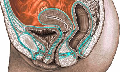
FEMALE = VAGINA, UTERUS, UTERINE TUBES, OVARIES

VAGINA- muscular tube lies posterior to bladder and anterior to the rectum.

It passes through both the UG and pelvic diaphragm.

UTERUS- know parts: cervix, isthmus, body, fundus

- uterine wall has 3 layers: perimetrium, myometrium, and endometrium

UTERINE TUBES = FALLOPIAN TUBE

  • - parts: fimbriae, infundibulum, ampulla, isthmus, intra-uterine (intramural) part
  • - ampulla is the normal site of fertilization
  • - Because the uterine tube is open to the peritoneal cavity, infections in the vagina can spread into the peritoneum causing peritonitis.

BROAD LIGAMENT= folds of peritoneum (the rain pouch Dr. Tucker wore) that suspend the uterus and allow it to move.

- In places, the broad lig. has tough connective tissue that, because anatomists have to name everything, have names:

Ligamentum Teres (Round Lig.)- from near the fundus of the uterus through the inguinal canal to the labia majora

Ovarian Lig.- connects the ovary to the uterus

Suspensory Lig. - from the ovary to the lateral wall of the pelvis; is a conduit for ovarian vessels

Mesosalpinx- part of broad lig. between the uterine tube and ovary

Mesovarium- where the ovary itself attaches to the broad lig.

Mesometrium- part of the broad ligament attached to the body of the uterus

Are you tired yet? Keep it up, youíre doing great.

PERINEUM

The anal canal and external genitalia make up the PERINEUM. The perineum is diamond shaped with the superior triangle being the urogenital triangle and the inferior triangle being the anal triangle.

anterior point: pubic symphysis

posterior point: tip of coccyx

lateral points: ishial tuberosities

The PERINEAL BODY is the tendinous center of the perineum and the point of attachment for many muscles (levator ani, transverse perineal, bulbospongiosus). It can be stretched or even torn during childbirth.

ANAL TRIANGLE = ANAL CANAL, RECTUM, ISCHIOANAL FOSSA, AND PUDENDAL CANAL

ANAL CANAL- blood and nerve supply differ on the two sides of the pectinate line


Blood supply

Venous Drainage

Nerve Supply

Above the Pectinate Line

Superior and Middle Rectal a.

Superior rectal v.

Pelvic splanchnic- muscular

Inf. hypogastric- sensory

Below the Pectinate Line

Middle and Inferior Rectal a.

Inferior rectal v.

Inf. rectal n from Pudendal n.

· Know the difference between internal and external hemorrhoids.

Internal hemorrhoids- dilations of the superior rectal v. and are not painful because pain is not sensed above the pectinate line.

External hemorrhoids-dilations of the inferior rectal v. and are painful because pain is sensed below the pectinate line.

RECTUM- a continuation of the sigmoid colon and makes a right angle at the puborectalis sling (formed by what muscle?)

  • - supplied by superior, middle, and inferior rectal arteries

ISHIOANAL FOSSA- filled with fat and connective tissue

  • - supports and allows the anal canal to expand during defecation
  • - is also a potential site for infection

PUDENDAL CANAL- fibrous tunnel found along the lateral wall of the ishioanal fossa

  • - carries the pudendal nerve, artery, and vein

MALE UROGENTIAL TRIANGLE

includes scrotum, penis, membranous urethra and superficial perineal muscles

PENIS : root, body, and glans

- made of corpus spongiosum (forms the glans and the bulb of the penis) and

corpus cavernosum (separates in the root to form two crura)

SUPERFICIAL PERINEAL MUSCLES= SUPERFICIAL TRANSVERSE PERINEAL M., BULBLOSPONGIOSUS M., ISCHIOCAVERNOSUS M.

- innervated by perineal n.

BULBOSPONGIOSUS M. - is assoc. with the bulb of the penis; when they contract urine, or semen is expelled from the urethra

In women, the bulbospongiousus m. acts as a vaginal sphincter.

ISHIOCAVERNOSUS M.- surrounds the crura and when it contracts, blood is forced from the crura into the body of the penis to help maintain erection

In women, the ishiocavernosus m. is involved in the erection of the clitoris.

FEMALE UROGENITAL TRIANGLE

VULVA = external female genitalia, includes: MONS PUBIS, LABIA MAJORA AND MINORA, VESTIBULE OF THE VAGINA, CLITORIS, BULB OF THE VESTIBULE, AND GREATER VESTIBULAR GLANDS

  • note that the scrotum is homologous to the labia majora
  • the labia minora cover the vestibule of the vagina
  • the clitoris is made of two corpora cavernosa that from crura

QUIZ TIME!!!!!!!!

1. Where does the obturator internus muscle exit out of?
2. What are the walls of the true pelvis?
3. What are the muscles of pelvic diaphragm?
4. What is the course of the pudendal nerve?
5. Does the obturator nerve come from the sacral plexus?
6. Name the parts of the broad ligament.
7. What is the blood supply above the pectinate line?
8. What does the corpus spongiosum form?

Wow you really are smart! Ready for more?

VERTEBRAL COLUMN: 33 vertebrae

7 cervical, 12 thoracic, 5 lumbar, 5 (fused) sacral, 4 (fused) coccygeal

  • the cervical and lumbar sections curve concave posteriorly
  • excessive lumbar curvature is called lordosis
  • the thoracic and sacrococcygeal parts curve concave anteriorly
  • excessive thoracic curvature is called kyphosis
  • make sure you can identify the body, arch, vertebral foramen, pedicle, lamina, transverse and spinal processes, and superior and inferior articular processes on a vertebrae

*** see your syllabus for distinguishing characteristics of each region***

  • - Intervertebral discs are between adjacent vertebral bodies and made of an annulus fibrosus surrounding the nucleus pulposus. They act as shock absorbers and if they bulge posteriorly they can compress spinal nerves. This is a common source of lower back and leg pain.
  • - A herniated disc is one where the annulus fibrosus weakens allowing the nucleus pulposus poke out and put pressure on a spinal nerve. This causes lots of lower back pain and is what clinicians call sciatica. ( Can you guess which nerve roots are pinched?)
  • - ATLANTOOCCIPITAL JOINT = C1 and occipital bone ® nodding head yes
  • - ATLANTOAXIAL JOINT = C1 and C2 ® shaking head no

BACK MUSCLES

EXTRINSIC- from vertebral column ® upper limb

- Superficial extrinsic back m.- help anchor the upper limb to the axial skeleton

include: traps, lats, rhomboids, and levator scapula

- Intermediate extrinsic back m.- accessory respiration m.

include: serratus posterior superior and inferior

INTRINSIC- completely assoc. with the vertebral column

  • - Superficial intrinsic back m.- SPLENIUS- rotate, extend and laterally flex the neck
  • - Intermed. intrinsic back m.- ERECTOR SPINAE- laterally flex and extend back
  • - Deep intrinsic back m.- TRANSVERSOSPINAL M.- rotate and stabilize the vertebral column

THORACIC WALL

RIBS - 12 total:

1-7 (sometimes 8) = TRUE ribs- directly attach at the sternum

(usu. 8), 9 and 10 = FALSE ribs- attach to the rib superiorly, not directly to the sternum

11and 12 = FLOATING ribs- do not attach at all to the sternum

STERNUM

3 parts: manubrium, body, and xiphoid (CPR landmark)

  • - superior part of manubrium = jugular notch
  • - manubriosternal joint = joint between manubrium and body, forms the sternal angle (landmark for 2d rib)

BREASTS- read more in your assigned reading

  • - mostly made of fat
  • - mammary glands which make milk can really only be seen when milk is being made (guys have mammary glands too!)
  • - The following arteries supply the breast: anterior intercostal branches of the internal thoracic a. (a branch of the subclavian a.), the lateral thoracic, thoracoacromial a, and posterior intercostal a.
  • - Most of the lymphatic drainage of the breasts is to the axillary lymph

INTERCOSTAL SPACE

THORACIC WALL MUSCLES- all innervated by intercostal n

  • External intercostal m.- elevates ribs
  • Internal intercostal m.- depresses ribs
  • Innermost intercostal m.-elevates ribs

- Between the internal and innermost m. are the intercostal vein, artery, and nerve. They run in or near the costal groove, or the inferior margin of the rib.

The intercostal nerves arise from the ventral rami of T1-T11

LUNGS

  • - Surrounded by a double layer of membrane called the PLUERA SAC
  • - inner layer of Pleura sac is the Visceral layer
  • - outer layer of Pleura sac is the Parietal layer
  • - The (potential) space between the two layers is called the Pleural cavity and if air fills this cavity (itís normally filled with pleural fluid) the lung will collapse.
  • - Know the regions of the pleura: mediastinal and diaphragmatic pleura and the costodiaphragmatic recess
  • -Know the parts of the lungs: apex, base, root, and hilum
  • -The root is the bundle of stuff (primary bronchi, pulmonary vessels, connective tissue, nerves, and lymphatics) that enter and exit the lung at the hilum.
  • - Left lung : oblique fissure ® superior and inferior lobes; cardiac notch
  • - Right lung: horizontal and oblique fissure ® superior, middle, and inferior lobes
  • -After entering the hilum, the primary bronchi branch sending a secondary bronchi to each lobe. The secondary bronchi branch into tertiary bronchi which supply a specific bronchopulmonary segment.
  • - Note that the right primary bronchus is shorter, wider, and runs more vertical than the left. This is why objects often get stuck in the right bronchus or right lung.
  • - Lungs receive parasympathetic innervation (by the VAGUS NERVE (cranial nerve X)), and sympathetic innervation.

The vagus n. sends fibers to the bronchoconstrictors and the sympathetic fibers innervate the bronchodilators.

PERICARDIUM AND HEART

PERICARDIUM = double walled fibrous sac around the heart

  • - FIBROUS LAYER- outer layer, maintains structure
  • - SEROUS LAYER- smooth, lubricated layer that allows for movement
  • - Parietal and Visceral layer and Pericardial cavity

BLOOD FLOW THROUGH THE HEART

De-Oxygenated blood:

In the superior and inferior vena cava ® RT. Atrium ® RT. Ventricle ® out the Pulmonary Arteries to the Lung

Oxygenated blood:

From the lungs into the Pulmonary veins to the LFT. Atrium ® LFT. Ventricle ® out the aorta to the body

STRUCTURES OF THE HEART

RIGHT ATRIUM
  • - Openings from the superior and inferior vena cava and the coronary sinus
  • - (Interatrial septum) Fossa ovalis- remenant of the foramen ovale in the fetus
LEFT ATRIUM
  • -4 pulmonary veins
VENTRICLES
  • - trabeculae carnae, atrioventricular valves (cusps, papillary muscles, chordae tendinae), semilunar valves
  • - The papillary muscles contract before the ventricles do. The papillary m. pull on the chordae tendinae to close the cusps and therefore close the valve to prevent blood from moving back into the atrium.

Right ventricle: Tricuspid and pulmonary valve

Left ventricle: Thicker because has to pump the blood against more ressistance (entire body vs. just to the lungs), bicuspid or mitral valve, and aortic valve

- When someone listens to your heartbeat, they are hearing the closing of valves.

Blood Supply of the Heart

- Blood needs to get to and from the heart itself. See pages 64 and 67 in your textbook.

  • Right coronay artery- originates in the aorta, runs in the coronary groove and gives off the right marginal branch and the posterior interventricular a.
    • - supplies the right atrium and right ventricle
  • Left coronay artery- originates in the aorta, gives off the circumflex branch and the anterior interventrical a.
    • - supplies the left atrium, left ventricle and interventricular septum
  • Anterior Cardiac vein drains directly into the right atrium.
  • All other cardiac veins drain into the coronary sinus of the right atrium.
  • Great cardiac vein- travels with the anterior interventricular a.
  • Middle cardiac vein- travels with the posterior interventricular a.
  • Small cardiac vein- travels with the marginal branch
  • -Look over your syllabus for clinical conditions
  • - The heart is innervated by vagus nerve and sympathetic trunk via the cardiac plexus.

CONDUCTING SYSTEM

- The heart has its own specialized conducting system in which impulses are conducted to pacemaker regions.

- The two atria contract together before the two ventricles contract (together).

  • SINOATRIAL NODE- pacemaker of the heart, located at the point where the superior vena cava enters the right atrium
  • ATRIOVENTRICULAR NODE- located above the coronary sinus in the atrial septum
  • ATRIOVENTRICULAR BUNDLE - Splits into right and left branches which end as Purkinje fibers, which cause ventricular contractions.

MEDIASTINUM

space between the two pleura sacs, does not include the lungs or pleura

Superior Mediastinum (from antr. to postr.)

  • -Thymus
  • - Brachiocephalic v, Superior Vena Cava, Aortic Arch
  • - Vagus Nerve (Cranial n. X)- passes posterior to root of lung
  • -Right vagus, left vagus, and recurrent laryngeal n.
  • - Phrenic- passes anterior to root of lung, supplies the diaphragm
  • -Trachea
  • -Esophagus- connects the pharynx to the stomach

Anterior Mediastinum

connective tissue, fat, and lymph nodes

Middle Mediastinum

pericardium and heart

Posterior Mediastinum

  • - Thoracic (Descending) aorta
  • - Thoracic Duct- receives lymph from left side of head, neck, and thorax, the left upper limb and the rest of the body below the diaphragm
  • - Azygous Vein- connects the superior and inferior vena cava
  • - Esophagus

QUIZ TIME!!!!!!!

1. How many vertebrae do you have?
2. How does a cervical vertebrae differ from a thoracic? From a Lumbar?
3. Which ribs are true? Which ones are false? Why?
4. What are some differences between the right and left lungs?
5. Do all arteries carry oxygenated blood?
6. What nerve innervates the diaphragm?
7. What is in the middle mediastinum? (No cheating, here!) Superior mediastinum? Posterior?


ABDOMINAL CAVITY REVIEW

I. Surface anatomy: xiphoid process, costal margin, anterior superior iliac spine, pubic crest, pubic tubercle, pubic symphysis, umbilicus, linea alba.

II. Muscles:

  1. External Oblique runs downward and forwards forming aponeurosis anteriorly, which forms inguinal ligament and the superfical inguinal ring of the inguinal canal.
  2. Internal Oblique is deep to the E. O. and runs upward.
  3. Tranversus abdominis is deep to the I. O. and runs horizontal. It comes together with I. O. to form conjoint tendon.
  4. Rectus abdominis runs along the linea alba (six packs)
  5. Tranversalis Fascia is a fascia between the abdominal muscles and the peritoneum.

III. Inguinal Canal - An obligue passage 3-5 cm long, through the abdominal wall; spermatic cord (Male) and round ligament (female) travel from the abdomen to the perineum. It has an arch form by the I. O. and Transversus abdominus. The abdominal open is the deep ring and opening to the strotum is the superficial ring.

Boundaries; anterior- E. O. and I. O. muscles, posterior - conjoint tendon and transversalis fascia, roof - arching of the I. O. and T. A. muscles. floor - inguinal ligament.

IV. Spermatic Cord: consists of ductus deferens, testicular a.(a branch off of abdomial aorta), internal spermatic fascia, Pampiniform Plexus (Varicocele - pooling of blood due to defective valves) , cremasteric fascia, and external spermatic fascia.

V. Scrotum: mostly skin and fascia; Tunica Vaginalis - a perioneal sac surrounding the testes (parietal layer and visceral layer). Hydrocele - accumulation of fluid in the cavity of the tunica vaginalis. Tunica albuginea - a tough fibrous capsule around the testes.

VI. What is the difference between Direct and Indirect Inguinal Hernia? know this....

Peritoneal Cavity, Esophagus and Stomach.

Remember that the human body is a 3D structure. So the peritoneum is like a balloon with its wall against the abdominal wall. Then structures such as the stomach and small intestine push though the wall and wrap them self in the peritoneal wall (like a fist punching through a balloon). These structures are suspended by the mesentery.

  1. Know the different between peritoneal (very mobile), retroperitoneal (partial cover by the peritoneal and not very mobile), and intraperitoneal structures (things inside the peritoneal cavity).
  2. Know the different between greater sac and lesser sac (omental foramen - an opening between greater and lesser sac). Greater omentum descends from the stomach and wraps back up to the transvers colon. Lesser omentum is from the live to the stomach.

Abdominal Viscera

  1. Esophagus - enters through the esophageal hiatus of the diaphragm. The distal end is where the lower esophageal sphinster locates. The opening to the stomach is the cardiac orifice.
  2. Stomach: (no digestion occurs here). Parts : cardiac part, fundus (mostly air), body, pyloric part (with the pyloric sphincter) and antrum (the site of gastrin secretion).
  3. Blood supply: Know this well... Celiac trunk comes off of the aorta. a, left gastric a. supplies esophagus and upper part of stomach (left of of lesser curvature). b, splenic a. gives off pancreatic branches, left gastroepiploic and short gastric a. (which supplies most of the greater curvature). c, Common hepatic - branches into hepatic a. proper and gastroduodenal artery. Hepatic artery proper gives off right gastric artery (supply the right side of lesser curvature) and branches into right and left hepatic arteries.
  4. What is Hiatus hernia? and Pyloric stenosis?

Small and Large Intestine

I. Duodenum (short but important structure); 1st part (upper duodenal cap) is free (peritoneal). 2nd (descending) recieved bile duct and pancreatic duct, which come together at the hepatonpancreatic ampulla, which opens at the major duodenal papilla. 3rd. (horizontal) - crosses anterior to the aorta. One other artery that cross in front of this part is the superior mesenteric a. 4th, ascending, connects with jejunum at the duodenojejunal flexure.

* Most (70-75 %) of ulcers occur at this structure.

II. Small intestine: jejunum (2/5) occupies the umbilical region and the ileum (3/5) occupies the hypogastric region. Blood supply by superior mesenteric a. and innervation is by the vagus and splanchnic nerves.

III. Large intestine: What are the differences between small instestine and large intestine? small intestine is usually very smooth. Large intestine has haustra, teniae coli, and appendices epiploicae.

  1. Cecum - is between small and large intestine. The open has a valve (iliocecal valve). What happens when feces plugged up the opening to the vermiform appendix?
  2. Ascending colon; retroperitoneal- not very mobile, blood supply right colic.
  3. Transverse colon - peritoneal - movable, supply by middle colic.
  4. Descending colon - retroperitoneal - not very mobile, supply by left colic.
  5. Sigmoid colon - look like and S. lies within the pelvis suspended from its wall by the sigmoid mesocolon. peritoneal - lots of movement.
  6. Innervation - parasympathetics by the vagus as far as the middle of the transverse colon.  Distal to that done by the pelvis splanchnics (S2-S4).  Sympathetic innervation via the thoracic splanchnic nerves and the collateral ganglia.

Abdominal Viscera

I. Liver

When you look at the liver in the text remember that the liver is flip up. Thus, you are looking at it posteriorly and upside-down. It has two surfaces (diaphragmic and visceral) and four lobes (right, left, quadrate (inferior to the caudate lobe), and caudate lobes.

  1. Ligaments: know the all, but major ones are falciform ligament and ligamentum teres.
  2. Function: protein synthesis, chemical modification, clotting factors, and important blood forming organ in fetus.
  3. Blood Supply: hepatic artery, proper , Make sure you know the Portal Vein system.

II. Gall Bladder: lies along the right edge of the quadrate lobe. The body drain into the cystic duct which combine with common hepatic duct to from bile duct. Bile duct joint with pancreatic duct to form hepatopancreatic ampulla which drains into the 2nd part of the duodenum.

  1. Function: to store bile for digesting fat.
  2. Blood supply: cystic a. and v.

III. The Pancreas: is an exocrine and endocrine gland consisting of a head, body, neck and tail. It is retroperitoneal which means it doesnít move that much. The pancreatic duct enters the duodenal papilla. Innervation is by sympathetic fibers parasympathetic fibers. Clinical conditions: pancreatitis and cancer of the pancreas.

IV. Spleen: is on the left side of the abdomen and posterior to the 9th and 10th costal margin. It is a lymphatic organ where antibody forms. There are two ligaments holding it up; gastrolienal which connects the spleen to the stomach and lienorenl which attaches the sleen to the posterior abdominal wall on the left side. Blood supply: Splenic a. from celiac trunk.

Anatomy Tables - Duodenum, Pancreas, Liver, & Gallbladder


Arteries

Artery

Source

Branches

Supply

Notes

aberrant hepatic

left - l. gastric a.; right- SMA


liver


celiac trunk

abdominal aorta at level of T12-L1 intervertebral disc

left gastric, splenic, common hepatic

stomach, lower esophagus, liver, upper duodenum, pancreas, spleen

(Greek, celiac = belly; the celiac artery supplies the principal organs of the belly)

cystic

right hepatic a. (or superior mesenteric a., proper hepatic a., left hepatic a., gastroduodenal a.)

no named branches

gall bladder

cystic a. arises from the right hepatic a. in ~72% of cases; other possible origins are noted at left and are clinically relevant during gall bladder surgery (Greek, cystic/kystis = bladder or pouch)

gastroduodenal

common hepatic

supraduodenal aa., retroduodenal aa., posterior superior pancreaticoduodenal a., anterior superior pancreaticoduodenal a., right gastro-omental a.

upper duodenum, upper part of head of pancreas; greater curvature of stomach on right


gastro-omental, left

splenic

gastric, omental

mid-part of greater curvature of stomach; greater omentum

anatomoses with right gastro-omental a.; also known as: left gastroepiploic a.

gastro-omental, right

gastroduodenal

gastric, omental

lower part of pyloric part of stomach, right part of greater curvature of stomach, greater omentum

anastomoses with left gastro-omental; also known as: right gastroepiploic a.

hepatic, common

celiac trunk

gastroduodenal & proper hepatic

liver & parts of duodenum, pancreas & stomach


hepatic, proper

common hepatic

right gastric, right & left hepatic

liver, lesser curvature of stomach


hepatic, left

proper hepatic


left & quadrate lobes of liver, part of caudate lobe

left hepatic a. supports liver parenchyma and stroma

hepatic, right

proper hepatic

cystic

right lobe of liver, part of caudate lobe

right hepatic a. supports liver parenchyma and stroma

splenic

celiac trunk

dorsal pancreatic, pancreatic brs., pancreatica magna, caudal pancreatic, short gastric, left gastro-omental, splenic brs.

pancreas, spleen, greater curvature of stomach


supraduodenal

gastroduodenal


1st part of duodenum superiorly


pancreatic, caudal

splenic


tail of pancreas


pancreatic, dorsal

splenic

right br., left br.

pancreas near neck

right br. anastomoses with anterior superior pancreaticoduodenal in prepancreatic arcade

pancreatic, inferior

dorsal pancreatic, left br.


lower part of body of pancreas


pancreatic, superior

splenic (multiple small brs.)


superior part of body of pancreas


pancreatica magna (great pancreatic)

splenic


body of pancreas


pancreaticoduodenal, anterior inferior

inferior pancreaticoduodenal, from superior mesenteric

pancreatic brs., duodenal brs.

lower duodendum & head of pancreas

anastomoses with anterior superior pancreaticoduodenal in anterior pancreatic arcade

pancreaticoduodenal, anterior superior

gastroduodenal

pancreatic brs., duodenal brs.

upper duodenum & head of pancreas

anastomoses with anterior inferior pancreaticoduodenal in anterior pancreatic arcade

pancreaticoduodenal, inferior

superior mesenteric

anterior & posterior inferior pancreaticoduodenal

lower duodenum & head of pancreas


pancreaticoduodenal, posterior inferior

inferior pancreaticoduodenal, from superior mesenteric

pancreatic brs., duodenal brs.

lower duodendum & head of pancreas

anastomoses with posterior superior pancreaticoduodenal in posterior pancreatic arcade

pancreaticoduodenal, posterior superior

gastroduodenal

pancreatic brs., duodenal brs.

upper duodenum & head of pancreas

spiral around common bile duct; anastomoses with posterior inferior pancreaticoduodenal in posterior pancreatic arcade


Veins

Vein

Tributaries

Drains Into

Region Drained

Notes

esophageal


azygos and hemiazygos vv.

esophagus

connect with esophageal brs. of left gastric, forming potential portal-caval anastomosis (esophageal varices when enlarged)

portal

formed by union of superior mesenteric & splenic vv.; tributaries: posterior superior pancreaticoduodenal v., right & left gastric vv.

right & left branches of portal v., into liver sinusoids


connects with caval drainage at 1) esophagus, 2) rectum, 3) umbilicus, 4) retroperitoneal gut structures

hepatic, left

segmental vv.

inferior vena cava

lateral segment & superior portion of medial segment of left lobe of liver


hepatic, middle

segmental vv.

inferior vena cava

inferior portion of medial segment of left lobe of liver, & inferior part of anterior segment of right lobe


hepatic, right

segmental vv.

inferior vena cava

posterior segment & superior portion of anterior segment of right lobe of liver


gastric, left

esophageal vv.

portal v.

lesser curvature of stomach, lower esophagus

connects with esophageal vv., forming portal-caval venous anastomosis (esophageal varices when enlarged); also known as: coronary v.

gastric, right


portal v.

lesser curvature of stomach

also known as: coronary v.

gastro-omental, left

gastric & omental brs.

splenic v.

greater curvature of stomach & greater omentum

also known as: left gastroepiploic v.

gastro-omental, right

gastric & omental brs.

superior mesenteric v.

greater curvature of stomach & greater omentum

also known as: right gastroepiploic v.

mesenteric, superior

pancreticoduodenal, jejunal, right and middle colic

portal v.

gut


pancreaticoduodenal, posterior superior


portal v.

posterior part of head of pancreas & 1st & 2nd part of duodenum posteriorly

connects with posterior inferior pancreaticoduodenal

pancreaticoduodenal, inferior

anterior & posterior inferior pancreaticoduodenal

superior mesenteric v.

lower part of duodenum & head of pancreas

connects with anterior & posterior superior pancreaticoduodenal vv.

pancreaticoduodenal, anterior superior


unites with right gastro-omental v. to drain to superior mesenteric

upper duodendum & head of pancreas anteriorly

connects with anterior inferior pancreaticoduodendal v.

paraumbilical


umbilical part of left branch of portal vein

falciform ligament, round ligament of liver

potential site of portal-caval anastomosis; blood may pass retrogradely into body wall veins near umbilicus, creating caput medusa sign

splenic

pancreatic brs.

unites with SMV to form portal v.

spleen


mesenteric, inferior

superior rectal, sigmoid, left colic vv.

splenic v. (or superior mesenteric v.)

gut from splenic flexure distally

courses lateral to 4th part of duodenum


Viscera

Organ

Location/Description

Notes

bile duct, common

union of cystic & common hepatic ducts

drains to hepatopancreatic ampulla & greater duodenal papilla with main pancreatic duct

duodenojejunal junction

suspended by suspensory muscle (ligament) of duodenum

inferior mesenteric v. passes to left side

intestine, small


3 parts: duodenum (1 foot long, mostly retroperitoneal); jejunim (8 feet long); ileum (12 feet long)

duodenum

C-shaped proximal segment of small intestine

4 parts: 1st or superior, 2nd or descending, 3rd or transverse, 4th or ascending (Latin, duodenum = 12, for it was estimated to be twelve finger-breadths long)

fusion fascia

a connective tissue plane in which the nerves and vessels of the descending colon reside


greater duodenal papilla

an eminence on the posteromedial wall of the descending duodenum

location of the opening of the hepatopancreatic duct (Latin, papilla = a nipple)

lesser duodenal papilla

located just superior to the greater duod. papilla

location of the opening of the accessory pancreatic duct (Latin, papilla = a nipple)

pancreas


functions as digestive & endocrine gland; main pancreatic duct drains tail, body & lower head to greater duodenal papilla within 2nd part of duodenum; accessory pancreatic duct drains upper body to lesser duodenal papilla

pancreas, body of

stretches to left from neck; continuous with tail

splenic a. lies on superior border, splenic v. lies behind body

pancreas, head of

right-most part

lies within C-shaped curve of duodenum

pancreas, neck of

junction between head & body extending to left

superior mesenteric a. arises behind neck

pancreas, tail of

left-most extent of pancreas, extending toward hilum of spleen within splenorenal ligament


pancreas, uncinate process of

lowest part of pancreas

tip of uncinate process lies behind superior mesenteric vessels

pancreatic duct, accessory

drains upper head of pancreas

opens onto lesser duodenal papilla

pancreatic duct, main

drains tail, body & lower head of pancreas

unites with common bile duct at hepatopancreatic ampulla

liver

consists of 4 anatomical lobes: left, right, quadrate & caudate

lies inferior to the diaphragm as high as 5th rib on right

bare area of liver

lies between anterior & posterior laminae of coronary lig.

in contact with diaphragm

porta hepatis

region of undersurface of liver where hepatic ducts, hepatic aa. & portal v. enter/leave


quadrate lobe

part of liver between bed of gall bladder & round ligament of liver

functionally part of left lobe (Latin, quadrate = square)

ligamentum venosum

lies in attachment of hepatogastric lig. to liver, between caudate & left lobe

remnant of obliterated ductus venosus

gallbladder

hangs below liver between right & quadrate lobes of liver

connected to common bile duct via cystic duct

gallbladder, body of

lies in bed of gall bladder between right & quadrate lobes of liver


gallbladder, fundus of

expanded lower part; located near inner surface of anterior abdominal wall at junction of right 9th costal cartilage & right semilunar line


neck, gallbladder

constricted upper part of gallbladder

continuous with cystic duct

hepatic duct, common

formed by union of right & left hepatic ducts

unites with cystic duct to form common bile duct

hepatic duct, left

drains left lobe, quadrate lobe & part of caudate lobe of the liver

unites with right hepatic duct to form common hepatic duct

hepatic duct, right

drains right lobe & part of caudate lobe of the liver

unites with left hepatic duct to form common hepatic duct

bile duct, common

union of cystic & common hepatic ducts

drains to hepatopancreatic ampulla & greater duodenal papilla with main pancreatic duct

cystic duct

duct of the gallbladder; unites with common hepatic duct to form common bile duct

both drains and fills gallbladder (Greek, cystic/kystis = bladder or pouch)


Peritoneal Specializations and Associated Structures

Specialization

Location/Description

Notes

circular folds of the pyloric sphincter

2nd part of the pyloric sphincter

more like the duodenum than the stomach

hepatoduodenal ligament

part of lesser omentum connecting liver with 1st part of duodenum

contains common bile duct, proper hepatic a. & portal v.

hepatogastric ligament

part of lesser omentum connecting liver with lesser curvature of stomach


falciform ligament

sickle-shaped fold of peritoneum connecting liver to umbilicus

contains round ligament of liver (ligament teres hepatis) (Latin and English combo, falciform = shaped like a scythe or sickle)

coronary ligament

peritoneum connecting liver to undersurface of diaphragm

area between anterior & posterior laminae is bare area of liver

pyloric sphincter

greatly thickened layer of the muscularis externa in the pylorus of the stomach

normally closed except when excreting chyme

round ligament of the liver


also known as: ligamentum teres hepatis

triangular ligaments

left and right extremes of the coronary ligament

formed by the fusion of anterior & posterior laminae of the coronary ligament

ligament of Treitz

posterior to the pancreas and splenic vein and anterior to the left renal vein

also known as the suspensory muscle of the duodenum

ligamentum teres hepatis

lies within falciform lig.; connects liver to umbilicus; remnant of umbilical v.

also known as: round ligament of liver (Latin, teres = round)

transverse mesocolon

peritoneum connecting transverse colon to posterior abdominal wall


mesentery

peritoneum connecting jejunum & ileum to posterior abdominal wall

also known as: mesentery of small intestine


Clinical Terms

Term

Definition

portocaval anastomosis

a common method for reducing portal hypertension by diverting blood from the portal venous system to the systemic venous system by creating a communication between the portal vein and the IVC. This usually leads to hepatic encephalopathy as the toxins from the GI tract bypass the liver and its processing of them.

pancreatitis

Acute or chronic inflammation of the pancreas, which may be asymptomatic or symptomatic and which is due to autodigestion of pancreatic tissue by its own enzymes. It is caused most often by alcoholism or biliary tract disease; less commonly it may be associated with hyperlipemia, hyperparathyroidism, abdominal trauma (accidental or operative injury), vasculitis or uremia.

pancreatic cancer

A malignant growth of the pancreas. More than 90% are ductal adenocarcinomas with islet cell carcinomas making up the difference. Symptoms include abdominal pain, abdominal swelling, jaundice, weight loss and anorexia.

gallstones

A concretion in the gall bladder or a bile duct, composed chiefly of cholesterol crystals.

jaundice

Yellowing of the skin (and whites of eyes) by bilirubin, a bile pigment. Frequently because of a liver problem. (Latin, jaundice/jaune = yellow)

cholecystectomy

The surgical removal of the gallbladder. This type of surgery may be performed with the traditional open incision or via a flexible fiberoptic scope (laparoscopic cholecystectomy).

cirrhosis

Liver disease characterized pathologically by loss of the normal microscopic lobular architecture, with fibrosis and nodular regeneration. The term is sometimes used to refer to chronic interstitial inflammation of any organ. (Greek, kirrhos = yellow liver + -osis = condition)

portal hypertension

Any increase in the portal vein pressure due to anatomic or functional obstruction (for example alcoholic cirrhosis) to blood flow in the portal venous system. Indicators of portal hypertension are: esophageal varices, hemorrhoids, enlarged veins on the anterior abdominal wall (caput Medusae) and ascites (fluid within the abdominal cavity).

ascites

An effusion and accumulation of serous fluid in the abdominal cavity. (Greek, askos = a bag)

Kocher manuever

Reflecting the duodenum and pancreas medially by cutting through the fusion fascia along the right side of the descending part of the duodenum. This maneuver is used to gain access behind the pancreas.

duodenal stenosis

Narrowing of the duodenum. May be congenital or the result of neoplastic growth. (Latin, stenosis = a narrowing)

duodenal atresia

Congenital duodenal atresia (blind end) of the duodenum, caused by a failure of recanalization of the duodenal lumen during development. It should be noted that 70% of patients with duodenal atresia also have other malformations including Down's Syndrome, cardiac or GI defects. (Greek, atresia = without hole)

cholecystojejunostomy

surgical establishment of a communication between the gall bladder and the jejunum.

pruritis

itching caused by drug reaction, food allergy, liver disease, cancers, parasites, aging or dry skin. (Latin, prurio = an itching)

bilirubin

pigment found in bile. Mainly formed during heme catabolism- the breakdown of old red blood cells within the liver. (Latin, bilis = bile + ruber = red)

hepatomegaly

enlargement of the liver

diuretics

agents that promote the excretion of urine through their effects on the function of the kidney

endoscopic retrograde cholangiopancreatography

fiberoptic endoscopy for duodenal observation and cannulation of vater's ampulla in order to visualize the pancreatic and biliary duct system by retrograde (against the usual direction of flow) injection of contrast media

hemangioma

an anomaly that leads to a mass that resembles neoplastic tissue. (Greek, -oma = tumor)

caput medusae

varicose veins radiating from the umbilicus. Ordinarily seen as a sign of cirrhosis of the liver and caused by portal hypertension. (Latin, caput medusae = medusa head)

cholecystitis

acute or chronic inflammation of the gall bladder

laparoscopic cholecystectomy

surgery to remove a diseased gallbladder through a fiberoptic scope that is inserted into a small incision near the navel. The patient is usually home 24 hours after the surgery.

hematemesis

vomiting of blood. (Greek, hema = blood + -emesis = vomiting)

Pringle maneuver

manually stopping the inflow to the liver by compressing the portal vein and proper hepatic artery. This is utilized in cases when the liver is bleeding and the bleeding must be stopped immediately (such as in trauma or surgical exploration)

·             Greater omentum

·             Gastrosplenic ligament

·             Splenorenal ligament

·             Gastrophrenic ligament

·             Gastrocolic ligament

·             Lesser omentum

·             Hepatogastric ligament

·             Hepatoduodenal ligament

·             Mesentery proper

·              

·             Ligaments and folds of peritoneum

·             Falciform ligament

·             Ligamentum teres hepatis (obliterated umbilical vein)

·             Coronary ligament

·             Left triangular ligament

·             Right triangular ligament

·              

·             Peritoneal cavity

·             Greater peritoneal sac

·             Omental (epiploic) foramen (of Winslow)

·             Lesser peritoneal sac (omental bursa)

·             INTERNAL FEATURES OF THE ANTEROLATERAL ABDOMINAL WALL

·             Umbilical folds

·             Median - contains the remnant of the urachus

·             Medial - contains the obliterated umbilical a.

·             Lateral - contains the inferior epigastric vessels

·              


          Abdomen: Peritoneum and Foregut


PERITONEUM

Definitions


Peritoneum--Mesothelial (Simple squamous epitheliums), glistening, transparent serous membrane lining the abdominal walls and contents

Parietal part--lines the abdominal and pelvic walls; receives same blood and nerve supply as the region it lines

Visceral part--lines the organs; receives same blood and nerve supply as organs

Peritoneal Cavity--The potential space of capillary thinness  between the parietal and visceral peritoneum; there are no organs in the peritoneal cavity, normally empty except for thin layer of fluid (peritoneal fluid) to keep surfaces moist, reduce friction, and supply leukocytes and antibodies; Subdivided into two sacs

Greater sac--entered by surgical incision of anterolateral abdominal wall; divided by transverse mesocolon into:

Supracolic compartment--contains the stomach, liver, and spleen

Infracolic compartment--contains the small intestine and ascending and descending colon; lies posterior to the greater omentum and is divided into right and left infracolic spaces by the mesentery of the small intestine

Lesser sac (omental bursa)--lies posterior to the stomach and adjoining structures and lesser omentum; permits free movement of the stomach on the structures posterior and inferior to it because wall slide smoothly.         

Superior recess--limited superiorly by the diaphragm and the posterior layers of the coronary ligament of the liver

Inferior recess--between the superior part of the layers of the greater omentum; mostly sealed off potential space

Peritoneal Reflections--peritoneum reflected from one area to another

(example--body wall to organ, organ to organ).  These reflections may be called omenta, mesenteries or ligaments.

Omentum--double-layered extension or fold of peritoneum that passes from the stomach and proximal part of the duodenum to adjacent organs in the abdominal cavity or to the abdominal wall

Mesentery--connection of viscera to body wall; double layered with thin layer of loose connective tissue between, in which vessels, nerves,  lymphatics and fat run; length of mesentery determines the degree of mobility

Peritoneal ligament--double layer of peritoneum that connects an organ with another organ or to the abdominal wall

Intraperitoneal--surrounded by peritoneum--inside the peritoneum; organ that is almost completely covered by peritoneum, except for the small area where the mesentery is attached (example transverse colon, stomach, spleen)

Retroperitoneal (extraperitoneal)--Behind the peritoneum--an organ that is covered with peritoneum only on its anterior surface (examples: duodenum and  pancreas [secondarily retroperitoneal], kidneys)

Embryology

Derivatives of the dorsal mesogastrium

Greater omentum--prominent peritoneal fold that hangs down like an apron from the greater curvature of the stomach and the proximal part of the duodenum; folds back and attaches to the anterior surface of the transverse colon and its mesentery; quite mobile (with peristalsis)

Functions--prevents the visceral peritoneum from adhering to the parietal peritoneum lining the abdominal wall; protects other viscera by walling-off an inflamed organ (like appendix) [abdominal policeman]; cushions organs against injury and forms insulation against loss of body heat

Closed sac in the male

Open sac in the female

Gastrophrenic ligament--connects the stomach to the inferior surface of the diaphragm

Gastrosplenic (gastrolienal) ligament--connects the stomach to the spleen; reflects to the hilum of the spleen

Gastrocolic ligament--connects stomach to transverse colon; apron-like part of greater omentum

Splenorenal ligament

Derivatives of the ventral mesogastrium

Lesser Omentum-helps former lesser sac, also called the omental bursa; cut off and isolated from the greater omentum due to the rotation of the stomach; opening into the lesser sac is called the epiploic foramen (aka omental foramen or foramen of Winslow; communication between greater and lesser sacs); connects the lesser curvature of the stomach and the proximal part of the duodenum to the liver

Hepatogastric (Gastrohepatic) ligament--membranous portion of lesser omentum connecting the liver to the stomach

Hepatoduodenal ligament--thickened free edge of the lesser omentum that conducts the portal triad (portal vein, hepatic artery, and bile duct)

Visceral peritoneum of the liver

Ligaments of the liver

falciform ligament--connects liver to the anterior abdominal wall

coronary ligament

triangular ligaments

Folds and Fossae (or recesses)--a recess (pouch) is formed by a peritoneal fold

Recto-uterine pouch (Pouch of Douglas)

Utero-visceral pouch

Hepatorenal pouch (“Morrison’s Pouch)

Paracolic gutters--communication between the supracolic and infracolic compartments; grooves between the lateral aspect of the ascending or descending colon and the posterolateral abdominal wall; provide pathways for the flow of ascitic fluid and the spread of infections

Anterior wall folds--A peritoneal fold is a reflection of peritoneum that is raised from the body wall by underlying blood vessels, ducts, and obliterated fetal vessels

Median--Urachus

Medial umbilical folds--Obliterated umbilical arteries

Lateral umbilical folds--Inferior epigastric arteries--remember their importance to defining inguinal hernia types

Blood Supply

Parietal peritoneum--from the abdominal wall

Visceral peritoneum--from vessels to the organs

Nerves

Parietal Peritoneum                                                                                                   Visceral Peritoneum

T7-L1                                                                                                                                                  T6-T12 thoracic splanchnics

Vasomotor from sympathetics                                                               Vasomotor from sympathetics

Pain: acute and localized                                                                                   Pain: Insensitive


FOREGUT

Parts

Arterial Supply

Celiac artery (“trunk”)--supplies the organs of the foregut and anastomoses with the superior mesenteric artery which supplies the midgut;  first unpaired branch of the abdominal aorta, is usually the second branch overall; many variations in branches

Left gastric artery--to stomach and esophagus--to lesser curvature of stomach and esophageal branch

Splenic artery--to spleen, stomach, and pancreas--short gastric arteries, left gastroepiploic artery, pancreatic arteries

Common hepatic artery--to liver, gallbladder, stomach, duodenum--

Proper hepatic artery--left hepatic artery, right hepatic artery, cystic artery, right gastric artery

Gastroduodenal artery--superior pancreaticoduodenal artery, right gastroepiploic artery

Venous Drainage

Gastric and splenic veins to the portal system

Innervation

Sympathetic--prevertebral [celiac] ganglia--causes vasoconstriction

Parasympathetic--vagus nerve--increases GI motility, secretion, absorption

Lymphatics--numerous, lymph nodes that surround each organ; all drain into the thoracic duct, cysterna chyli

Stomach

Orifices

Cardial-physiologic sphincter (not anatomical, no muscle band)

Pyloric --guarded by pylorus (pyloric sphincter), first sphincter in GI tract ”gatekeeper”; normally in tonic contraction and is closed except when emitting chyme; gastric peristalsis passes chyme into small intestine

Curvatures

Lesser--shorter, concave border; angular notch (incisure) is sharp indentation approximately 2/3 of the distance along the lesser curvature that indicates the junction of the body and the pyloric part; attachment of lesser omentum

Greater--attachment of greater omentum--longer, convex border

Parts

Cardia--part surround the cardial orifice

Fundus--air bubble-looks black in radiographs; dilated superior part that relates to the left dome of diaphragm; limited by the plane of the cardial orifice; superior part reaches level of left 5th intercostal space; cardial notch is between esophagus and fundus; may be dilated by gas, fluid, food

Body--lies between the fundus and the pylori antrum

Pyloric (antrum, canal, pyloris)--funnel-shaped region; wide part (pyloric antrum) leads to pyloric canal (narrow part); pyloric sphincter controls discharge of the stomach contents through the pyloric orifice into duodenum

Relations

Anterior--adjacent to left lobe of  liver, diaphragm, anterior abdominal wall

Posterior-- omental bursa and pancreas

Bed of stomach--posterior wall of omental bursa (diaphragm, spleen, kidney, suprarenal gland, pancreas, splenic artery and mesocolon

Muscle coats

Longitudinal

Circular

Oblique

Interior--

Gastric folds (rugae, longitudinal ridges) form when gastric mucosa is contracted, most marked in pyloric part

Gastric canal--forms temporarily during swallowing between gastric folds along lesser curvature; saliva and small amounts of food pass through to pyloric canal

Arteries--come off of three main branches of celiac trunk

Left gastric--branch off celiac trunk; runs in lesser omentum to the cardia and then courses along the lesser curvature to anastomose with the right gastric

Right gastric--arises from hepatic artery; runs to the left along the lesser curvature to anastomose with the left gastric

Gastroduodenal

Right Gastroepiploic (gastro-omental)--arises as one of two terminal branches of the gastroduodenal artery; runs to left along greater curvature and anastomoses with left gastroepiploic

Left gastroepiploic (gastro-omental)--arises from the splenic artery and courses along greater curvature to anastomose with the right gastroepiploic           

Short gastric (4 or 5)--arise from distal end of splenic artery or its splenic branches and pass to the fundus of the stomach

Veins--parallel arteries in position and course and drain into portal system

Left and right gastricčPortal vein

Short gastric, left gastroepiploic and pancreaticčsplenic vein, which joins superior mesenteric veinčportal vein

right gastroepiploic and pancreaticčSuperior mesenteric vein

prepyloric--čright gastric vein; used to identify pylorus

Lymphatics--accompany arteries along greater and lesser curvatures and drain into gastric and gastro-omental lymph nodes located in the curvaturesčceliac lymph nodes

Nerves--

Parasympathetic--from anterior (derived from left vagus) and posterior (derived mainly from right vagus)  vagal trunks

Sympathetic--from T6-T9 passes to celiac plexus through the greater splanchnic nerve

Applied Anatomy

Hiatal hernia--protrusion of stomach into the mediastinum through the esophageal hiatus; two types--sliding hiatus, cardial portion through diaphragm and paraesophageal hiatus, fundus part through with cardial portion undisturbed

Peptic ulcer--lesions of the stomach commonly associated with the presence of Heliobacter pylori; an ulcer is formed when the mucosa is inadequate to protect the mucosa from erosion by acid

Gastroscopy--use of flexible fiber endoscopes to examine the mucosa of stomach to observe gastric lesions and take biopsies

Gastrectomy--removal of all or part of the stomach in cases of stomach carcinoma or peptic ulcer disease

Vagotomy--section of the vagal trunks at the esophageal hiatus to reduce the production of acid

Duodenum--first, shortest, widest and most fixed part of small intestine; C-shaped course around the head of the pancreas from right to left;

4 Parts--First (proximal) two parts derived from embryological foregut and principally supplied by celiac artery (by definition are foregut), second (distal) two parts derived from embryological midgut and principally supplied by mesenteric artery (are midgut by definition)

Ampulla--first 2 cm of superior part--next to pylorus; is mobile and has different appearance than rest of duodenum

Part I (superior)--short (5 cm); lies anterolateral to the body of L1 vertebra--ascends from the pylorus and is overlapped by the liver and gallbladder; only has peritoneum anteriorly (except ampulla); proximal part has the hepatoduodenal ligament attached superiorly and the greater omentum attached inferiorly

Anterior: Liver, neck of the gall bladder

Posterior: Gastroduodenal artery, common bile duct, and portal vine

Superior: neck of gall bladder

Inferior: pancreas

Part II (vertical or descending)--longer (7-10 cm); descends along the right sides of L1-L3; runs inferiorly, curving around head of pancreas; retroperitoneal

Anterior: Liver, transverse colon, jejunum, ileum, gallbladder

Posterior: Right kidney, ureter, renal blood vessels

Medial: Head of pancreas (pancreatic duct), common bile duct--enter posteromedial wall and untie to form the hepatopancreatic ampulla which opens on an eminences located posteromedially in the descending duodenum, the major duodenal papilla

Lateral: right kidney

Part III (horizontal or inferior) (6-8 cm); runs transversely to the left, passing over the IVC, aorta and L3 vertebra; it is crossed by the superior mesenteric artery (SMA) and vein and the root of the mesentery of the jejunum and ileum

Anterior: superior mesenteric vessels, root of mesentery, transverse colon and mesocolon

Posterior: right psoas muscle, IVC, aorta, sympathetic trunks

Superior:   pancreas

Inferior: Coils of jejunum and ileum

Part IV (Ascending) short (5 cm) begins left of L3 and rises to superior of L2; curves anteriorly at border of body of pancreas to join the jejunum at the duodenojejunal junction that takes the form of an acute angle, the duodenojejunal flexure which is supported by a suspensory muscle of the duodenum

Anterior: Root of mesentery and coils of jejunum

Posterior: Left psoas muscle

Suspensory ligament of the duodenum (“Ligament of Treitz”)

Interior Features

Plicae cicularis

Duodenal papilla--hill on side through which ducts open

Major papilla--combine pancreas and biliary tree ducts

Minor papilla--pancreatic duct only

Opening of main pancreatic duct (“Ampulla of Vater”)

Opening of accessory pancreatic duct

Arteries--the duodenal arteries arise from the celiac trunk and the superior mesenteric artery

Celiac trunk č gastroduodenal artery č superior pancreatico-duodenal arteries--supplies duodenum proximal to the entry of the bile duct into the second part of the duodenum

Superior mesenteric arteryčinferior pancreaticoduodenal artery--supplies duodenum distal to the entry of the bile duct

Both pancreaticoduodenal arteries lie in the curve between the duodenum and the head of the pancreas and supply both structures.  Their anastomosis, near the level of entry of the bile duct is in effect an anastomosis of the celiac and superior mesenteric artery.

Veins--follow arteries and drain into the portal vein, either directly or through the superior mesenteric and splenic veins

Lymphatics--follow the arteries; anterior vessels drain into pancreaticoduodenal lymph nodes and pylori lymph nodes; posterior vessels drain into superior mesenteric lymph nodes; efferent vessels drain into celiac lymph nodes

Innervation--From vagus and sympathetic nerves through the celiac and superior mesenteric plexuses on the pancreaticoduodenal arteries

Duodenal recesses

Superior

Inferior

Paraduodenal--large and lie to left of ascending part of duodenum; may strangulate a loop of intestine (paraduodenal hernia)

Retroduodenal

Applied Anatomy

Ulcers--normally in the posterior wall of the superior part of the duodenum; close relation also makes it possible to effect liver and gallbladder

Atresia

Stenosis


Liver--RUQ (mainly) and LUQ, largest gland in the body; protected by rib cage a little under sternum; stores glycogen and secretes bile

Surface projections

Surfaces--diaphragmatic and visceral; diaphragmatic separated from diaphragm by subphrenic recesses which are separated into right and left by falciform ligament

Anterior: anterior abdominal wall

Posterior: diaphragm, inferior vena cava, “bare area”

Superior: diaphragm

Inferior (visceral): right kidney, colon, stomach, duodenum, right suprarenal gland, lesser omentum

Lobes--are demarcated by 4 furrows which make an H-shape; independent right and left lobes, each with own bloody supply and biliary drainage and demarcated from one another by grooves formed by gallbladder (inferiorly) and the IVC (superiorly)

Right--attachment of falciform ligament

Left--includes caudate lobe and much of quadrate lobe

Quadrate--between gallbladder and falciform ligament (posterior surface)

Caudate--between inferior vena cava and ligamentum venosum (posterior)

Portal triad--portal vein, hepatic duct, hepatic artery proper are enclosed in thick free edge of the lesser omentum, specifically the hepatoduodenal ligament

Peritoneal “Ligaments”

Coronary

Falciform

Triangular

Arteries--30% of total hepatic blood supply

Common hepatic artery--from the celiac trunk to the origin of the gastroduodenal artery

Proper hepatic artery--from the origin of the gastroduodenal artery to its bifurcation into right and left branches

Right and Left hepatic arteries--supply right and left lobes; carries well-oxygenated blood from aorta (right supplies middle part of bile duct)

Veins--70% of total hepatic blood supply

Portal vein--carries poorly oxygenated but nutrient-rich blood from the GI tract to the sinusoids of the liver

Hepatic veins--intersegmental in their distribution and function, draining parts of adjacent segments; formed by the union of the central veins of the liver;  open into the IVC, just inferior to diaphragm

Gallbladder--repository for cholesterol which is necessary to digest fat; bile salts emulsify to be carried through blood; bile is made in the liver and constantly dribbles down bile duct; it then flows retrogradely into gallbladder to sit until fatty meal is eaten; lies in gallbladder fossa on the visceral surface of the liver, which is at the junction of the right and left lobes of the liver

Surface projection--tip of 9th costal cartilage on the right side; first part of duodenum is usually stained with bile; capacity of up 50 mL of bile.

Parts

Fundus--wide end, projects from the inferior border of the liver and is usually located at the tip of the 9th costal cartilage in the MCL

Body--contacts the visceral surface of the liver, the transverse colon, and the superior part of the duodenum

Neck--narrow, tapered and directed toward the porta hepatis; mucosa of neck spirals into a fold, the spiral valve;

Cystic duct--connects the neck of gallbladder to the common hepatic duct; passes between the layers of the lesser omentum, usually parallel to the common hepatic duct, which it joins to form the bile duct

Arteries

Cystic artery--supplies gallbladder, cystic duct and proximal part of the bile duct; commonly arises from right hepatic artery in the angle between the common hepatic duct and the cystic duct

Hepatic artery

Veins--cystic veins--drain directly into the liver via the bed of the gallbladder and the portal system

Biliary Tree

Bile Passages--Common hepatic duct and cystic duct unite to form the common bile duct, which empties into the Sphincter of Oddi

Bile Ducts--right and left hepatic ducts drain right and left lobes of liver, respectively; they then unite to form the common hepatic duct, which is joined on the right side by the cystic duct to form the bile duct, which conveys bile to the duodenum

Bile Duct--forms in the free edge of the lesser omentum by the union of the cystic duct and common hepatic duct; descends posterior to first part of duodenum and lies in a groove on the posterior surface of the head of the pancreas; comes in contact with main pancreatic duct on left side of descending duodenum and unite to form the hepatopancreatic ampulla (ampulla of Vater), with the dilation within the major duodenal papilla; circular muscle around distal end of duct is thickened to form the sphincter of the bile duct which when contracted doesn’t allow bile to enter duodenum and backs up into the cystic duct to the gallbladder where it is concentrated and stored

Applied anatomy of liver and gallbladder

Jaundice

Gallstones

Variations in gall bladder anatomy

Pancreas--an elongated, accessory digestive gland

Surface projection

Parts

Head--expanded part of the gland; nestled in C-curve of duodenum, firmly attached to medial aspect of descending and horizontal part of the duodenum; rests posteriorly on IVC, right renal artery and vein, left renal vein

 uncinate process--a projection from the inferior part of the head, extends medially to the left, posterior to the SMA

Bile duct--lies in a groove on the posterosuperior surface of the head or is embedded in its substance

Neck--short and overlies superior mesenteric vessels, which form a groove in its posterior aspect; anterior surface is adjacent to the pylorus of stomach

Body--lies to left of SMV and SMA, passing over aorta and L2, posterior to the omental bursa; anterior surface lies in floor of omental bursa and forms part of stomach bed; posterior surface devoid of peritoneum and contacts aorta, SMA, left suprarenal gland, left kidney and renal vessels

Tail--continues to spleen; lies anterior to left kidney, closely related to hilum of spleen and left colic flexure; relatively mobile and passes through layers of splenorenal ligament with the splenic vessels


Relations

Anterior: transverse mesocolon; small gut, stomach

Posterior: splenic vein, aorta, IVC, bile duct

Right: duodenum

Left: spleen

Ducts--possibly two entries into duodenum

Main pancreatic duct--mainly dorsal; beings in tail and runs through parenchyma to the head where it turns inferiorly and is closely related to the bile duct; unites with bile duct to form short hepatopancreatic ampulla which opens into the descending part of the duodenum at the major duodenal papilla.  Sphincters (sphincter of pancreatic duct, sphincter of bile duct and hepatopancreatic sphincter of Oddi) are smooth muscles sphincters that control the flow of bile and pancreatic juice into the duodenum

Accessory pancreatic duct--drains uncinate process and inferior part of head and opens into duodenum at minor duodenal papilla; usually communicates with main pancreatic duct

Arteries

Splenic--form several arcades with pancreatic branches of gastroduodenal and superior mesenteric arteries--supply body and tail

Superior pancreaticoduodenal--branches of gastroduodenal--supply head

Anterior and posterior inferior pancreaticoduodenal--branches of SMA supply head

Veins--corresponding pancreatic veins are tributaries of splenic and superior mesenteric part os the portal vein, but most drain into splenic vein

Spleen--mobile, largest lymphatic organ; usually purplish color; located intraperitoneally in LUQ; completely surrounded by peritoneum except at hilum where veins and arteries enter and leave; varies considerably in size, weight and shape but usually size of clenched fist

Surface projections--separated from ribs by diaphragm and costodiaphragmatic recess

Upper border--related to the 9th rib

Lower border--related to the 11th rib

Relations

Posteriorly: left part of diaphragm

Anteriorly:  Stomach

Medially--Kidney and tail of pancreas (left boundary of omental bursa)

Inferiorly--left colic flexure


“Ligament” contents and relationships

Lienorenal ligament--splenic artery and tail of pancreas

Gastrolienal (gastrosplenic) ligament--connects spleen to greater curvature of stomach--short gastric artery and stomach

Phrenicocolic ligament--diaphragm

Arteries--Splenic artery, follows tortuous course along superior border of pancreas; between layers of splenorenal ligament divides into five or more branches that enter the hilum

Veins--Splenic vein, formed by several tributaries that emerge from the hilum; joined by IMV and runs posterior to the body and tail of the pancreas throughout most of its course ; joins with SMV to form portal vein


For the following questions, indicate the letter that corresponds to the SINGLE MOST APPROPRIATE ANSWER.

1. Dr. Nussbaum used a laparoscopic procedure to remove the gallbladder of a 53-year-old
woman. He knew that all of the following statements concerning the gallbladder are
correct EXCEPT that it
    A. is located between the right and quadrate lobes of the liver.
*  B. is commonly supplied by visceral afferent fibers that traverse the superior
        mesenteric plexus.
    C. has a duct that joins the common hepatic duct.
    D. receives arterial supply via a vessel that usually branches from the right hepatic
        artery.
    E. has a surface that may contact the anterior body wall.

2. During vaginal delivery, an episiotomy is performed by incising the perineal body to
prevent uncontrolled tearing of the perineum. All of the following muscles are attached
to the perineal body EXCEPT the
    A. bulbospongiosus.
    B. deep transverse perineal.
    C. external anal sphincter.
    D. superficial transverse perineal.
*  E. ischiocavernosus.

3. A 57-year-old woman has a tumor involving the labia majora and minora. Surgery is
recommended to remove not only the tumor, but also any affected lymph nodes. To
which lymph nodes would tumor cells most likely initially metastasize?
    A. Common iliac
    B. Inferior mesenteric
    C. Lumbar
    D. Sacral
*  E. Superficial inguinal

4. A 67-year-old man is jaundiced and has severe abdominal pain. He is diagnosed with
cancer of the head of the pancreas. What spinal cord segment(s) receive the visceral
afferent fibers from the head of the pancreas?
    A. T1 to T4
*  B. T5 to T9
    C. T10 and T11
    D. T12
    E. L1 and L2

5. During an emergency hysterectomy in a 45-year-old woman, the surgeon exposes a
uterine artery prior to its ligation. What structure passing inferior to this vessel and
lateral to the cervix must be protected from accidental ligation during this procedure?
    A. Fallopian tube
    B. Ovarian artery
*  C. Ureter
    D. Pudendal nerve
    E. Ovarian ligament

6. A 26-year-old woman complains of abdominal pain in the umbilical region. A problem
with her appendix is suspected. Through which of the following nerves would visceral
afferent fibers from the appendix travel?
    A. Vagus
    B. Greater splanchnic
*  C. Lesser splanchnic
    D. Least splanchnic
    E. Lumbar splanchnic

7. A 12-year-old boy is undergoing liver transplant surgery. During the procedure the
structure immediately posterior to the epiploic foramen of Winslow is damaged causing a
significant amount of bleeding into the operative field. What vessel has most likely been
injured?
    A. Aorta
    B. Common hepatic artery
*  C. Inferior vena cava
    D. Portal vein
    E. Right renal vein

8. A 63-year-old man had a tumor adjacent to the celiac ganglion and plexus. This tumor
would probably compress all of the following EXCEPT
    A. sympathetic preganglionic fibers.
*  B. parasympathetic postganglionic fibers.
    C. visceral afferents from the stomach.
    D. parasympathetic preganglionic fibers.
    E. sympathetic postganglionic fibers.

9. A 63-year-old woman has an ulcer in the posterior wall of the first part of the duodenum.
Which of the following structures is most likely to be directly damaged if a perforation
occurred at the site of the ulcer?
    A. Right suprarenal gland
    B Ascending colon
    C. Aorta
*  D. Gastroduodenal artery
    E. Superior mesenteric vein

10. An aortic aneurysm in the aortic hiatus of the diaphragm could directly compress the
    A. vagal trunks.
    B. lesser splanchnic nerves.
*  C. azygos vein.
    D. right phrenic nerve.
    E. sympathetic chains.

11. A 55-year-old man has cancer of the stomach requiring a total gastrectomy. Which group
of lymph nodes should be examined as a possible site of initial metastasis?
    A. Superior mesenteric
    B. Lumbar
    C. Internal iliac
    D. Inferior mesenteric
*  E. Celiac

12. During the examination of a 45-year-old woman, a large pelvic mass is discovered.
After determining that it is an ovarian tumor, surgery is scheduled. During the surgical
procedure, the primary vascular supply to the ovary must be ligated. Clamping and
cutting which of the following ligaments will accomplish this task?
*  A. Suspensory
    B. Round
    C. Ovarian
    D. Cardinal
    E. Broad

13. The T8 spinal nerve is compressed by a tumor in the intervertebral foramen. This
compression could result in decrease or loss of innervation to all of the following
structures EXCEPT
    A. a portion of the transversus abdominis muscle.
    B. a portion of the rectus abdominis muscle.
    C. postganglionic cell bodies in the celiac ganglion.
    D. skin of the anterior abdominal wall above the umbilicus.
*  E. the cremaster muscle.

14. A 23-year-old motorcycle accident victim was rushed to the operating room, where a
large volume of blood was aspirated (removed) from his abdominal cavity. The bleeding
resulted from tearing of the ileocolic artery near its origin. This vessel was ligated to
prevent further blood loss. The cecum in this patient continued to receive adequate blood
flow by way of direct communication of branches of the ileocolic with those of the
    A. jejunal arteries.
    B. cecal arteries.
*  C. right colic artery.
    D. left colic artery.
    E. appendicular artery.

15. In a patient with severe portal venous obstruction secondary to cirrhosis of the liver,
blood containing nutrients absorbed from the jejunum would most likely reach which of
the following organs first?
    A. Pancreas
    B. Spleen
    C. Lung
    D. Ileum
*  E. Heart

16. A 45-year-old man arrived at the emergency room with a severe pain on his right side.
He said the pain first started just below his ribs. Now, five hours later, the pain has
moved to his groin. Suspecting that the patient is passing a ureteric calculus (stone), the
physician ordered an intravenous pyelogram (IVP). In interpreting the pyelogram, she
recalled that all of the following statements about the ureter are correct EXCEPT that
*  A. at the hilus of the kidney, the pelvis of the ureter is the most anterior structure.
    B. the ureter has several constrictions along its course where a calculus
        may pass with difficulty.
    C. it runs vertically downward posterior to the parietal peritoneum and lies on the
        psoas muscle.
    D. it enters the pelvic cavity by crossing anterior to the bifurcation of the common
        iliac artery.
    E. pain from the pelvic portion of the ureter is attributed to afferent fibers which
        travel in the lumbar splanchnic nerves.

17. A 37-year-old male was diagnosed with testicular cancer. Cells in a tumor arising in the
testes would directly metastasize to the
    A. superficial inguinal lymph nodes.
*  B. lumbar lymph nodes.
    C. external iliac lymph nodes.
    D. internal iliac lymph nodes.
    E. intestinal lymph nodes.

18. A fourth-year medical student assists the surgeon in a direct inguinal hernia repair of a
60-year-old man. This type of hernia
    A. is due to a congenital defect.
*  B. can pass through the superficial inguinal ring.
    C. is not surrounded by peritoneum.
    D. courses through the deep inguinal ring.
    E. passes posterior to the inguinal ligament.

19. Hemorrhoids bother a 23-year-old woman who is seven months pregnant. These have
developed due to the enlargement of the normal anastomotic connections between
branches of the inferior mesenteric vein and branches of which of the following veins?
*  A. Internal pudendal
    B. Obturator
    C. Esophageal
    D. Paraumbilical
    E. Uterine

20. The urologist palpated the patient's spermatic cord while performing a vasectomy. He
knew that all of the following structures were within at least two of the coverings of the
spermatic cord EXCEPT the
    A. ductus deferens.
    B. testicular artery.
    C. pampiniform plexus of veins.
*  D. ilioinguinal nerve.
    E. testicular lymphatics.

21. A 46-year-old man is having an inguinal hernia repaired. During the procedure the
superficial inguinal ring must be enlarged to reduce the hernia and retract the intestinal
loop. Which of the following structures is being incised to accomplish this task?
    A. Conjoined tendon
    B. Rectus sheath
    C. Transversalis fascia
    D. Internal oblique muscle
*  E. External oblique aponeurosis

22. During abdominal surgery, the surgeon examined several structures that are primarily
(not secondarily) retroperitoneal. These structures included all of the following EXCEPT
the
    A. kidneys.
*  B. pancreas.
    C. obliterated allantois.
    D. bladder.
    E. suprarenal glands.

23. A 22-year-old man is shot in a bar fight. The bullet passes through the midline of the
anterior abdominal wall immediately superior to the pubic symphysis. If the bullet
continued in a horizontal direction, in the mid-sagittal plane, which of the following
structures would most likely be penetrated?
    A. Abdominal aorta
    B. Internal iliac artery
    C. Ureter
    D. Spinal cord
*  E. Urinary bladder

24. In preparation for removal of an inflamed gallbladder, the surgeon laparoscopically
examined the cystic duct and the common bile duct and confirmed that the common bile
duct usually lies
*  A. anterior to the portal vein.
    B. posterior to the inferior vena cava.
    C. lateral to the descending portion of the duodenum.
    D. to the left of the proper hepatic artery.
    E. posterior to the pancreas.

25. A 52-year-old man complains of pain in the epigastric region. This is most likely
referred pain resulting from which of the following?
    A. Rectal polyps
    B. Renal calculus
    C. Testicular tumor
    D. Transverse colon obstruction
*  E. Duodenal ulcer

26. A patient with a massive carcinoma of the stomach had most of his stomach removed
surgically (subtotal gastrectomy). In that procedure, vessels that may be ligated
USUALLY include branches or tributaries of all of the following EXCEPT the
    A. splenic artery.
*  B. superior mesenteric artery.
    C. proper hepatic artery.
    D. gastroduodenal artery.
    E. portal vein.

27. A 68-year-old woman has a recently diagnosed occlusion of her inferior mesenteric artery
near its origin. If significant collateral circulation fails to develop, she will have an
increased risk for ischemia in which of the following parts of her gastrointestinal tract?
    A. Stomach
    B. Ileum
    C. Transverse colon
*  D. Sigmoid colon
    E. Anal canal

28. In discussing the problems linked with alcoholism to a patient who has begun drinking
three glasses of wine per day to avoid heart disease, you remind him that alcohol absorbed from the jejunum goes directly to the liver, by a short route involving the
    A. splenic vein.
    B. left gastric vein.
    C. right gastric vein.
*  D. superior mesenteric vein.
    E. left gastroepiploic vein.

29. A 56-year-old man is having surgery to repair a direct inguinal hernia. The surgeon is
using an open procedure during which a nerve is damaged. The man awakens after
surgery and finds that he has lost sensation in the anterior part of the scrotum and the
medial part of the adjacent thigh. What nerve has most likely been injured?
    A. Iliohypogastric
*  B. Ilioinguinal
    C. Lateral femoral cutaneous
    D. Pudendal
    E. Subcostal

30. A 63-year-old man has his rectum removed after a diagnosis of rectal cancer. Following
this procedure he is unable to have an erection. Along with this deficit, what portion of
the gastrointestinal system would most likely lose its parasympathetic innervation?
    A. Stomach
    B. Duodenum
    C. Ileum
    D. Ascending colon
*  E. Descending colon

31. A 67-year-old woman is having reconstructive surgery to correct prolapse of the uterus.
During this procedure, manipulation of which of the following ligaments could cause
injury to the uterine vessels?
*  A. Cardinal (transverse cervical)
    B. Ovarian
    C. Round
    D. Suspensory
    E. Uterosacral

32. A 36-year-old man is passing blood in his urine. A pyelogram reveals an abnormality in
the portion of the collecting system immediately following the major calyces. Where is
the abnormality located?
    A. Renal cortex
    B. Renal medulla
    C. Minor calyx
*  D. Renal pelvis
    E. Ureter

33. An obese, 45-year-old woman has severe upper abdominal pain after eating a meal at a
fast food restaurant. She is diagnosed as having a large number of stones in her
gallbladder. What nerves carry the visceral afferent fibers involved in the transmission of
"pain" from the gallbladder?
*  A. Greater splanchnic
    B. Lesser splanchnic
    C. Lumbar splanchnic
    D. Pelvic splanchnic
    E. Vagus

34. A 42-year-old man is brought to the emergency department with a severe laceration of
the anus. Branches of which of the following structures carry pain fibers from this
region?
    A. Pelvic splanchnic nerves
    B. Sacral splanchnic nerves
*  C. Pudendal nerves
    D. Inferior hypogastric plexus
    E. Sympathetic chains

35. During repair of an inguinal hernia, the inferior epigastric artery was cut as it passed
close to the deep inguinal ring. If this vessel were ligated, there would be a
compensatory increase in blood flow through the superior epigastric artery, which is located
    A. between the internal oblique and transversus abdominis muscles.
    B. in the subserous fascia deep to the rectus sheath.
*  C. between the rectus abdominis muscle and the posterior layer of the rectus sheath.
    D. deep to the body of the sternum.
    E. in the fatty layer of the abdominal superficial fascia.

36. A 47-year-old man is diagnosed with a hiatal hernia. During the surgical repair of this
hernia, the vagus nerves are damaged and determined to be non-functional. Which of
the following abdominal organs would still have their parasympathetic nerve supply intact?
    A. Stomach
    B. Jejunum
    C. Ileum
    D. Ascending colon
*  E. Sigmoid colon

37. As part of a weight therapy program, a 35-year-old man underwent resection of
portions of his greater omentum. During the procedure, his surgeon recalled that the
greater omentum
    A. contains the left gastric artery.
    B. is made up of two peritoneal layers.
    C. is attached to the lesser curvature of the stomach.
    D. is attached to the lower border of the pancreas.
*  E. contains the left gastroepiploic artery.

38. A 47-year-old man with abdominal pain has an abdominal CT scan. It reveals an
obstruction of the third part of the duodenum due to an arterial aneurysm. In what vessel
is the aneurysm most likely located?
    A. Celiac artery
    B. Gastroduodenal artery
    C. Inferior mesenteric artery
    D. Right renal artery
E. Superior mesenteric artery

39. After birth, the vessel(s) that bring blood from the placenta to the heart of the embryo
close off. A postnatal remnant of this system is the
    A. urachus.
    B. median umbilical ligament.
    C. medial umbilical ligament.
    D. lateral umbilical ligament.
*  E. ligamentum teres (round ligament of the liver).

40. While exploring the posterior abdominal wall you recall that all of the following relations
of the lumbar spinal nerves are correct EXCEPT that the
    A. lateral femoral cutaneous nerve passes deep to the inguinal ligament.
*  B. lumbosacral trunk is located lateral to the psoas major muscle.
    C. genitofemoral nerve is usually located on the anterior surface of the psoas major
        muscle.
    D. obturator nerve is located medial to the psoas major muscle.
    E. ilioinguinal nerve passes through the superficial inguinal ring.

41. An infant arrives in the emergency department with symptoms of gastrointestinal
obstruction including abdominal distention and tenderness. Ultrasound reveals that part
of the intestine is twisted around a ligament connecting the ileum to the umbilicus. This
omphalomesenteric ligament is a rudiment of which of the following structures?
    A. Allantois
    B. Median umbilical ligament
*  C. Vitelline duct
    D. Gubernaculum
    E. Hepatoduodenal ligament

42. Each of the following structures is involved in the development of the diaphragm
EXCEPT the
    A. septum transversum.
    B. pleuroperitoneal membrane.
    C. body wall.
    D. esophageal mesenchyme.
*  E. pleuropericardial folds.

43. Ligaments derived from the ventral mesentery include all of the following EXCEPT the
    A. coronary.
    B. falciform.
*  C. gastrosplenic.
    D. hepatoduodenal.
    E. hepatogastric.

44. An infant is born with a rare defect at the umbilicus which expresses urine. It is probably
a(n)
    A. omphalocele.
    B. Meckel's diverticulum.
    C. omphalomesenteric fistula.
    D. patent vitelline duct.
*  E. urachal fistula.

45. An infant in your care is diagnosed with a malformation of the GI tract. MRI and barium
radiography indicate that the gut is organized in such a way that the midgut viscera are in
their normal locations in the peritoneal cavity but the duodenum lies anterior to the
transverse colon. You conclude that abnormal rotations of the gut must have occurred
resulting in a total rotation of the intestines of
*  A. 90 degrees clockwise.
    B. 270 degrees counterclockwise.
    C. 270 degrees clockwise.
    D. 90 degrees counterclockwise.
    E. 180 degrees clockwise.

46. The former location of the membrane separating the endodermal and ectodermal portions
of the anorectal canal is marked in the adult by an irregular folding of mucosa called the
    A. cloaca.
    B. urogenital sinus.
*  C. pectinate line.
    D. proctodeum.
    E. urorectal septum.

47. An infant has a 46,XY karyotype but exhibits a normal female phenotype (ovaries,
female genital ducts and external genitalia). During development there was the
disruption of expression of
    A. androgen receptors.
    B. 5-(-reductase.
*  C. TDF.
    D. anti-Mullerian hormone.
    E. a hormone produced by the Leydig cells.

48. Both mesonephric ducts sprout two ureteric buds which bifurcate but fail to penetrate the
metanephric blastema. This would result in
*  A. oligohydramnios.
    B. polycystic kidney disease.
    C. formation of nephric (renal vesicles).
    D. a bifid ureter.
    E. hydronephrosis.

49. An infant is born with female external genitalia and undescended testes within the
inguinal canals. It is subsequently determined that this baby has a disabling mutation of
the enzyme 5-(-reductase. All of the following internal genital structures are most likely
missing or undifferentiated in this infant EXCEPT the
    A. prostate gland.
    B. superior end of the vagina.
    C. uterus.
*  D. epididymis.
    E. testis cords.

50. Decreased amniotic fluid volume in the absence of causative factors (such as the rupture
of fetal membranes) should alert the sonographer (ultrasound technician) to all of the
following abnormalities EXCEPT
    A. pulmonary hypoplasia.
    B. bilateral renal agenesis.
    C. Potter's syndrome.
    D. limb deformities.
*  E. a rectourethral fistula.
1
 I. FOR THE FOLLOWING QUESTIONS, INDICATE THE LETTER THAT
CORRESPONDS TO THE SINGLE MOST APPROPRIATE ANSWER.

1. The trachea
    A. lacks diffuse lymphoid tissue (BALT).
    B. has a wall that includes elastic cartilage.
*  C. contains abundant longitudinally oriented elastic fibers.
    D. is surrounded by a complete ring of smooth muscle.
    E. is lined with stratified squamous epithelium.

2. Sebaceous glands
    A. secrete their product by the apocrine mode.
    B. secrete their product into eccrine gland ducts.
    C. secrete their product with the aid of myoepithelial cells.
*  D. include differentiating cells in which SER is abundant.
    E. can be classified as simple tubular glands.

3. The osmolarity of (provisional) urine is not appreciably altered once it
    A. leaves the proximal convoluted tubule.
    B. leaves the distal convoluted tubule.
    C. enters the distal convoluted tubule.
    D. enters Bowman's urinary space.
*  E. leaves the papillary ducts.

4. A recent article in the New England Journal of Medicine documented the use of an inhibitor of
angiotensin converting enzyme (ACE) to lower blood pressure and thereby reduce the incidence
of heart attack and stroke. This inhibitor would be most active directly on the
    A. macula densa.
    B. adrenal gland.
    C. glomerulus.
*  D. lung endothelial cells.
    E. juxtaglomerular cells.

5. Which statement is correct?
*  A. Renal medullary rays are cortical columns of straight ducts and tubules.
    B. The periphery of a renal lobule is defined by interlobar arteries.
    C. The pars convoluta lacks connecting tubules.
    D. The lumen of the renal pelvis is lined by simple cuboidal epithelium.
    E. Renal columns consist of the papillary collecting ducts.

6. Products in provisional urine that are reabsorbed by cortical nephrons enter the
interstitium where they are removed by the
    A. descending thick limbs.
    B. collecting ducts.
    C. vasa rectae.
    D. medullary rays.
*  E. peritubular capillary plexus.

7. In normal tissue, which of the following cell types would you expect to have the fewest
lysosomes?
    A. Osteoclasts
*  B. Schwann cells
    C. Neutrophils
    D. Promyelocytes
    E. Dust cells

8. Which of the following cell types lack a basal lamina or external lamina?
    A. Columnar cells of respiratory epithelium
    B. Smooth muscle cells
*  C. Fibroblasts
    D. Endothelial cells
    E. Podocytes

9. Which type(s) of muscle has/have one synapse on each cell?
*  A. Skeletal muscle
    B. Cardiac muscle
    C. Smooth muscle
    D. Smooth and cardiac muscle
    E. Cardiac and skeletal muscle

10. A mutant mouse lacks the enzyme that phosphorylates acid hydrolases on mannose residues at
position 6. A major function of which component of the kidney would be most adversely
affected?
    A. Distal convoluted tubules
    B. Collecting ducts
    C. Thin descending limb
*  D. Proximal convoluted tubules
    E. Juxtaglomerular cells

11. Under normal conditions, lamellar bodies or their products may appear in which of the
following cell types?
    A. Type I pneumocytes only
    B. Type II pneumocytes only
    C. Alveolar macrophages only
    D. Type I pneumocytes and alveolar macrophages
*  E. Type II pneumocytes and alveolar macrophages

12. Which region of the human respiratory tract would have small plates of hyaline cartilage and
bundles of smooth muscle?
*  A. Bronchus
    B. Bronchiole
    C. Alveolus
    D. True vocal cord
    E. Concha

13. The biochemical composition of surfactant within alveoli most closely resembles
*  A. low density lipoprotein (LDL).
    B. glycophorin.
    C. type I collagen.
    D. glycosaminoglycans.
    E. fibronectin.

14. The thinnest regions of a Type I pneumocyte measure
    A. (10 nm.
*  B. 25-50 nm.
    C. 100-250 nm.
    D. 500-1000 nm.
    E. (1000 nm.

15. The total number of renal lobules is identical to the number of
    A. renal pyramids.
*  B. medullary rays.
    C. interlobular arteries.
    D. minor calyces.
    E. renal corpuscles.

16. Within the juxtaglomerular apparatus renin is secreted by the
    A. macula densa.
*  B. modified arteriolar smooth muscle cell.
    C. modified arteriolar endothelial cell.
    D. specialized fibroblast.
    E. extraglomerular mesangial cell.

17. The formation of hypertonic urine in humans is possible because
    A. the urethra is lined by stratified squamous epithelium.
*  B. juxtamedullary nephrons extend into the medulla.
    C. blood remains isotonic throughout the vasa rectae.
    D. collecting duct permeability to water increases as ADH concentration decreases.
    E. the majority of renal corpuscles are in the inner cortex.

18. Which of the following regions would be most intensely stained by antibodies specific for a
surface marker on Clara cells?
    A. Nasal epithelium
    B. Tracheal epithelium
*  C. Respiratory bronchioles
    D. Interstitial spaces
    E. Alveoli

19. A sagittal (longitudinal) section through the epiglottis was compared to several cross sections cut
through the upper half of the structure. One of the main differences observed was that only the
sagittal section included
    A. elastic cartilage.
    B. stratified squamous epithelium.
*  C. pseudostratified ciliated columnar epithelium with goblet cells.
    D. mucous glands.
    E. diffuse lymphoid tissue.

20. Regarding the nasal cavity, which of the following is correct?
    A. The superior concha is responsible for most of the humidification of inspired air.
    B. The primary location for the olfactory epithelium is the inferior concha.
    C. Mucous, but not serous glands, are found within the lamina propria of the conchae.
    D. The epithelium of the superior concha is primarily non-keratinized stratified squamous.
*  E. The shape of the conchae is maintained primarily by the supportive bones that they
         contain.

21. Human olfactory epithelium includes
    A. ciliated supportive cells.
*  B. neurons that are regularly renewed.
    C. cells with receptors located on microvilli.
    D. basal cells with myelinated axons.
    E. a stratified squamous epithelium.

22. The Langerhans cells of skin
    A. are derived from the embryonic neural crest.
*  B. contain Birbeck granules that are in continuity with the plasma membrane.
    C. are attached to keratinocytes by desmosomes.
    D. are found primarily in the stratum basale.
    E. present, but do not process antigens.

23. Cells derived from a serous gland of a mutant mouse were grown in culture. The secretory
product synthesized by the cells was isolated from the culture medium and analyzed for its
protein and carbohydrate content. The only abnormality in the isolated glycoprotein was the
absence of sialic acid. This result led the investigator to suspect that there were defective
enzymes in the
    A. smooth endoplasmic reticulum.
    B. rough endoplasmic reticulum.
*  C. Golgi apparatus.
    D. secretory vesicles.
    E. signal recognition particles.

24. Alport's syndrome results from defective glomerular filtration caused by an inherited defect in
    A. proteoglycan aggregates.
    B. reticular fibers.
*  C. type IV collagen.
    D. lamin.
    E. fibrillin.

II. IN THE FOLLOWING SECTION, NOTE THAT THE CORRECT ANSWER IS THE
STATEMENT THAT IS LEAST LIKELY.

25. Regarding the vasa rectae, the following statements are true EXCEPT they
 * A. form the boundary between renal lobules.
    B. are oriented in the same direction as the thin limbs of the loops of Henle.
    C. drain into arcuate veins.
    D. arise from efferent arterioles of juxtamedullary nephrons.
    E. supply blood to the medulla.

26. In which compartment would you LEAST expect to find the protein laminin?
    A. Rough endoplasmic reticulum
    B. Cis-Golgi apparatus
    C. Trans-Golgi apparatus
*  D. Peroxisomes
    E. The extracellular matrix

27. The following structures are derived from matrix cells of the hair follicle EXCEPT
    A. medulla of hair.
    B. cortex of hair.
    C. cuticle of hair.
    D. internal root sheath.
*  E. external root sheath.

28. Each of the different segments of bronchioles includes all of the following components EXCEPT
*  A. goblet cells.
    B. ciliated cells.
    C. smooth muscle.
    D. Clara cells.
    E. elastic fibers.

29. Regarding the renal corpuscle, all of the following statements are correct EXCEPT
    A. the basal lamina (basement membrane) of the filtration barrier is formed by both
        podocytes and endothelial cells.
    B. the filtration slit diaphragms extend between pedicels from adjacent podocytes.
    C. fluid in the glomerular capillaries filters through fenestrations that lack diaphragms.
*  D. glucose passes through the filtration barrier, but is mostly reabsorbed in the distal
        convoluted tubule.
    E. mesangial cells may be adjacent to endothelial cells.

30. Which of the following is/are LEAST likely to be a component of the blood/air barrier?
    A. Type I cells
    B. Fused basal laminae
*  C. Smooth muscle cells
    D. Surfactant
    E. Endothelial cells

31. The following cell types typically may have more than one nucleus EXCEPT
    A. skeletal muscle.
    B. surface cell of transitional epithelium.
    C. osteoclast.
*  D. megakaryocyte.
    E. giant cells.

32. All of the following statements are true for the pulmonary arteries EXCEPT they
    A. carry deoxygenated blood.
    B. follow the bronchiolar tree.
    C. give rise to the alveolar capillary beds that surround alveoli.
    D. are elastic arteries.
*  E. are found in septa between lobules.

33. Within the integument, both the stratum basale and stratum spinosum are found in all of the
following EXCEPT
    A. external root sheath.
*  B. internal root sheath.
    C. nail bed.
    D. thin skin.
    E. thick skin.

34. One hour prior to sacrifice, a radioactive DNA precursor was injected into a young mouse.
Subsequently, a sagittal (longitudinal) section of the entire mouse was prepared for
autoradiography. Which of the following cell types is LEAST likely to show labeling?
    A. Chondrocytes of the epiphyseal plate
    B. Neutrophilic myelocyte
    C. Keratinocyte in stratum basale
*  D. Neuron in dorsal root ganglion
    E. Type II pneumocyte

III. A SET OF LETTERED HEADINGS IS FOLLOWED BY A LIST OF WORDS OR PHRASES. FOR EACH NUMBERED WORD OR PHRASE INDICATE THE CORRESPONDING LETTER.
    A. Membrane coating granules
    B. Keratohyalin granules
    C. Melanosomes
    D. A and B
    E. A and C

35. Originate in cell derived from neural crest
C

36. Lack a unit membrane
B

37. Product interacts with intermediate filaments
B

38. Product forms intercellular moisture barrier.
A

39. Minimal in cells below level of stratum spinosum.
D

A. Apocrine sweat glands
B. Eccrine sweat glands
C. Sebaceous glands
D. Apocrine and eccrine sweat glands
E. Eccrine and sebaceous glands

I. FOR THE FOLLOWING QUESTIONS, INDICATE THE LETTER THAT CORRESPONDS TO THE SINGLE MOST APPROPRIATE ANSWER.

1. Which of the following cell types is/are capable of division?
    A. Myelocyte
    B. Multipotential stem cell
    C. Orthochromatophilic erythroblast
*  D. A and B
    E. A, B and C

2. Which hematocrit and reticulocyte count combination is most likely for a Cincinnati resident, after three days of skiing in Vail, Colorado (high altitude)?
    A. 60% hematocrit, 0.8% reticulocytes
    B. 60% hematocrit, 4% reticulocytes
    C. 40% hematocrit, 0.8% reticulocytes
*  D. 40% hematocrit, 4% reticulocytes
    E. 20% hematocrit, 20% reticulocytes

3. Which of the following produce(s) mature circulating membrane-bounded elements that are
anucleate?
    A. Megakaryocyte
    B. Proerythroblast
    C. Lymphoblast
*  D. A and B
    E. A, B and C

4. Which statement below is true?
    A. Yellow marrow is so named because it is the site of white blood cell formation.
    B. Specific granules of eosinophils are highly acidophilic due to their content of
        heparin.
    C. Monocytes characteristically have a round, heterochromatic nucleus.
*  D. Azurophilic granules are found in platelets.
    E. Megakaryocytes are large, multinucleated cells.

5. Secondary (specific) granules first appear in which stage of granulocyte
development?
    A. Myeloblast
    B. Promyelocyte
*  C. Myelocyte
    D. Metamyelocyte
    E. Band cell

6. The appearance of multiple nucleoli in a proerythroblast is indicative of
    A. polyploidy.
*  B. transcription of clusters of ribosomal RNA genes.
    C. G2 arrest.
    D. a plane-of-section artifact.
    E. pluripotency.

7. An electron micrograph of a small blood vessel in cross section shows several types of
cells. One cell type is found between the lumen of the blood vessel and a wavy band
of acellular material that stains very lightly. Higher magnification views of the same
blood vessel show that these cells are joined together by tight junctions. These cells are
    A. macrophages.
    B. fibroblasts.
    C. pericytes.
*  D. endothelial cells.
    E. smooth muscle cells.

8. "Anchoring filaments"
    A. insert into desmosomes of intercalated discs.
*  B. provide structural support for certain lymphatic vessels.
    C. are impulse conducting elements found in the myocardium.
    D. are found in the adventitia of large blood vessels.
    E. are cytoskeletal elements.

9. Which of the following substances is not present in normal lymph?
    A. Serum immunoglobulins
    B. Serum electrolytes
*  C. Hemoglobin
    D. Peptide hormones
    E. Growth factors

10. In the formation of an atherosclerotic plaque, platelet derived growth factor (PDGF)
plays which of the following dual roles? It
    A.enhances adhesion of platelets and stimulates proliferation of smooth muscle
        cells.
    B. enhances adhesion of platelets and stimulates lipid uptake by monocytes.
    C. stimulates both lipid uptake by monocytes and proliferation of smooth muscle cells.
    D. attracts smooth muscle cells to the plaque and enhances differentiation of mono-
         cytes to macrophages.
*  E. attracts smooth muscle cells to the plaque and stimulates their proliferaton.

11. Which of the following are enclosed by a connective tissue capsule and have both
afferent and efferent lymphatics?
    A. Lymph nodes, Peyer's patches and thymus
    B. Lymph nodes and Peyer's patches
    C. Lymph nodes and thymus
    D. Peyer's patches and thymus
*  E. Lymph nodes

12. The predominant cell type in the lymph node paracortex and in internodular areas of lymphoid
        tissue is
    A. B-cells.
*  B. T-cells.
    C. plasma cells.
    D. dendritic cells.
    E. macrophages.

13. In an individual born with agammaglobulinemia (i.e., the inability to mount antibody
mediated immune responses), which would not occur?
    A. Allograft rejection
    B. Contact sensitivity
    C. Graft versus host disease
*  D. Immediate hypersensitivity
    E. Phagocytosis

14. A twenty-nine year old female presents with a thymoma (a malignant tumor of the thymus
gland). Following surgical removal of the thymus, which would be expected?
    A. A gradual loss of the ability to mount an immune response
    B. A marked reduction in the levels of serum IgG
    C. Loss of immunological memory
    D. Disappearance of T-cells in the peripheral blood
*  E. Little to no effect on the immune status

15. Which of the following functions in an autocrine mode during the maturation of T-helper precursor cells to committed T-helper cells?
    A. Interleukin-1
*  B. Interleukin-2
    C. Interleukin-6
    D. GM-CSF
    E. Erythropoietin

16. Proteolytic cleavage of an intact protein antigen into several antigenic peptides occurs
    A. at the cell surface.
*  B. in the endosomal compartment.
    C. in the lysosomal compartment.
    D. in the cisternae of the Golgi apparatus.
    E. in the cisternae of the rough endoplsmic reticulum.

17. A 5-year-old patient presents with recurrent viral infections. Subsequent tests reveal normal
levels of serum IgM and IgG. Additionally, the patient was immunized against polio virus as an
infant and still has normal levels of circulating antibody against the virus. Based on this information, the most likely diagnosis is
    A. plasma cell defect.
    B. macrophage defect.
    C. defect in antigen processing and presentation.
*  D. T-cell defect.
    E. B-cell defect.

18. Platelets
*  A. may be found in a storage pool.
    B. are not involved in clot retraction.
    C. have a peripheral region comprised primarily of intermediate filaments.
    D. survive approximately 200 days in circulation.
    E. have a central, clear hyalomere.

19. Which of the following are properties of mature red blood cells? They
    A. have eosinophilic staining properties because of their high content of mitochondria.
*  B. are about the same diameter as small lymphocyte nuclei in smears.
    C. are normally found at a concentration of about 5 x 10⁵ µl of blood.
    D. are phagocytic cells.
    E. freely enter connective tissue through continuous blood vessel walls.

Use the following experimental model to answer the next three questions (20-22).
About 15-20% of humans with severe chronic neutropenia have a genetic mutation that results in abnormal granulocyte colony stimulating factor receptors (GCSFR). Transgene technology was used to produce a mouse model in order to fully analyze the effects of the mutant GCSFR gene, designated as ?715. Mice with the following genotypes were available for study: normal wildtype (wt/wt); and heterozygous (wt/?715) and homozygous (?715/?715) mutants. The formed elements of peripheral blood (graph below) and bone marrow (table below) for all genotypes were evaluated.

20. Based on the data given in the graph on the left, which conclusions are justified?
*  A. Neutrophils are the predominant cell affected by the (715 GCSFR mutation.
    B. Megakaryocytes are likely to be affected.
    C. Only homozygous ((715/(715) mutants are affected.
    D. A and B
    E. A, B and C

21. CFU-NM are unlikely to be affected by the (715 GCSFR mutation because
    A. GCSF is known to act only at very late stages of hemopoeisis.
*  B. monocyte levels are normal.
    C. platelet levels are normal.
    D. other than neutrophils granulocyte levels are normal.
    E. each colony stimulating factor (CSF) affects only a single cell lineage.

22. Using the information in both the graph and table, which step in the differentiation of
neutrophils is strongly affected by the (715 GCSFR mutation?
    A. band form ==> segmented from
    B. metamyelocyte ==> band form
    C. myeloblast ==> promyelocyte
*  D. mature neutrophils in bone marrow ==> circulating neutrophils
    E. myelocyte ==> metamyelocyte

23. Which of the following responses to a foreign body is most closely associated with neutrophils?
    A. Anaphylaxis
    B. Antigen presentation
    C. Immediate hypersensitivity
*  D. Phagocytosis
    E. Allograft rejection

24. The destruction of lymphocytes within the thymus occurs by
    A. an autoimmune process.
*  B. apoptosis.
    C. necrosis.
    D. antibody mediated cytotoxicity.
    E. an inflammatory process.

25. In order for cytotoxic T-lymphocytes to eliminate virus-infected cells, which combination of Major Histocompatibility Complex (MHC) factors is required for cytotoxic T-cell activation and subsequent target cell killing?
activation of cytotoxic T-cells target cell killing
    A. class I MHC class I MHC
    B. class I MHC class II MHC
*  C. class II MHC class I MHC
    D. class II MHC class II MHC
    E. class II MHC MHC not required

II. IN THE FOLLOWING SECTION, NOTE THAT THE CORRECT ANSWER IS THE STATEMENT THAT IS LEAST LIKELY.

26. Which of the following is NOT a characteristic of pluripotential hemopoietic stem cells?
    A. Transplantable
    B. Capable of self-replication
    C. Capable of differentiating into more than one type of cell
    D. May be found in peripheral blood
*  E. Morphologically distinguishable in bone marrow smears

27. Cell proliferation is a property of the following cells associated with the adult circulatory
system EXCEPT
    A. endothelial cells.
    B. pericytes.
    C. vascular smooth muscle cells.
*  D. cardiac muscle cells.
    E. fibroblasts.

28. All of the following statements regarding leukocyte granules are correct EXCEPT
    A. specific granules of neutrophils are smaller than azurophilic granules.
    B. in the mature neutrophil, the specific granules are more numerous than the primary
        granules.
    C. basophil granules contain histamine.
*  D. eosinophilic and neutrophilic specific granules are equal in size, but differ in
        staining properties.
    E. neutrophils have bacteriocidal enzymes present in their granules.

29. Characteristics of white blood cells include the following EXCEPT they
    A. exhibit chemotaxis.
    B. exhibit diapedesis.
*  C. have density greater than red blood cells.
    D. have life spans from days to years.
    E. function in connective tissue.

30. Which of the following is LEAST likely to be present in the light region of
a germinal center?
*  A. Epithelial reticular cells
    B. Macrophages
    C. Mitotic cells
    D. Large lymphocytes
    E. Small lymphocytes

III. MATCH THE NUMBERED QUESTIONS BELOW WITH THE LETTERED OPTIONS A-E. EACH ANSWER MAY BE USED ONCE, MORE THAN ONCE OR NOT AT ALL.

Match the lettered options below with the numbered descriptive phrases related to the
heart.
    A. Endocardium
    B. Myocardium
    C. Annulus fibrosis
    D. Purkinje fibers
    E. None of the above

31. Continuous with the tunica intima of blood vessels
A

32. Thick in the ventricle and thinner in the atrium
B

Match each numbered description with the appropriate lettered vessel below.

A. Lymph capillary
B. Blood capillary
C. Muscular artery
D. Elastic artery
E. Large vein

33. Vessel may have associated pericytes
B

34. Has muscular adventitia that is thick
E

Match the lettered options below with the numbered descriptive phrases related to the
lymphoid system.

A. Lymph node only
B. Spleen only
C. Thymus only
D. Lymph node and spleen
E. Lymph node and thymus

35. Nodules in cortex
A

36. Subcapsular sinuses present
A

37. Medulla present
E

38. Supportive stromal cells derived from endoderm
C

III. DIAGRAMS

Use the SEM image from a lymphoid organ below to answer questions 39-40.

39. What structure is marked by X's?
    A. Trabecular vein
    B. Penicillar arteriole
*  C. Venous sinusoid
    D. Central artery
    E. Continuous capillary

40. What structure is shown at Y?
    A. Trabecula
    B. Connective tissue capsule
    C. White pulp
*  D. Cord of Billroth
    E. Germinal center

I. FOR THE FOLLOWING QUESTIONS, INDICATE THE LETTER THAT
CORRESPONDS TO THE SINGLE MOST APPROPRIATE ANSWER

1. Parenchymal cells of which of the following can be regarded as modified sympathetic
postganglionic neurons that have lost their axons and dendrites?
    A. Thyroid
    B. Parathyroid
*  C. Adrenal medulla
    D. Adrenal cortex
    E. Pineal

2. The primary secretory product from which of the following pancreatic cells is rich in
bicarbonate?
*  A. Duct cells
    B. Islet endocrine cells
    C. Exocrine acinar cells
    D. Islet capillary endothelial cells
    E. Exocrine pancreas capillary endothelial cells

3. A cross section from near the tip of the tongue would include
    A. taste buds on filiform papillae.
    B. skeletal muscle organized as inner circular and outer longitudinal layers.
    C. circumvallate papillae.
*  D. taste buds on fungiform papillae.
    E. papillae on both ventral and dorsal surfaces of the tongue.

4. The mediastinum testis originates from the
*  A. tunica albuginea.
    B. parietal layer of the tunica vaginalis.
    C. dermis of the scrotum.
    D. spermatic cord.
    E. interstitium of the seminiferous tubules.

Large amounts of colloidal material are characteristically found in the
    A. pars nervosa.
    B. pars distalis.
    C. pars tuberalis.
*  D. pars intermedia.
    E. infundibulum.

6. The histologist determined that a section was NOT from the esophagus because of the presence of
    A. a muscularis mucosae.
*  B. serous glands in the submucosa.
    C. stratified squamous epithelium that was not keratinized.
    D. diffuse lymphoid tissue in the lamina propria.
    E. skeletal muscle that was oriented both circularly and longitudinally.

7. Spermatozoa first acquire motility in the
    A. ampulla of the seminal vesicle.
    B. colliculus seminalis.
    C. ductus deferens.
*  D. epididymis.
    E. vagina.

8. The seminal vesicle
    A. stores the majority of mature spermatozoa.
    B. is composed of 30 to 50 tubuloalveolar glands.
*  C. requires testosterone for maintenance and activity.
    D. secretes a mucous lubricant during sexual arousal.
    E. has three well-defined layers of smooth muscle.

9. The portal tract (portal canal, portal triad) may include one or more branches of
    A. bile duct, portal vein, hepatic artery, central vein.
*  B. portal vein, hepatic artery, lymphatic, bile duct.
    C. portal vein, hepatic artery, sublobular vein, bile duct.
    D. hepatic vein, hepatic artery, bile duct, lymphatic.
    E. portal vein, hepatic vein, bile duct, lymphatic.

10. Lipid storage droplets characteristically are observed in the cytoplasm of
    A. Sertoli cells.
    B. peritubular cells.
    C. primary spermatocytes.
    D. secondary spermatocytes.
*  E. interstitial cells of Leydig.

11. Analysis of sections of developing teeth reveals that
    A. cementum is the major constituent of the tooth.
    B. odontoblast cell bodies occupy lacunae in dentin.
    C. hollow tubes containing cell processes extend through the width of the enamel.
    D. ameloblasts are found within the pulp cavity of the permanent tooth.
*  E. the enamel organ initially is connected to the dental lamina.

12. Which of the following secretes a slightly acidic fluid that is rich in acid phosphatase?
    A. Rete testes
*  B. Prostate gland
    C. Ductus deferens
    D. Seminal vesicles
    E. Bulbourethral gland

13. Which of the following indicates a correct order of structures encountered from the center
to the outside edge of an ovarian follicle? (Some structures in each sequence may be
missing.)
    A. Zona pellucida®theca interna®basement membrane®theca externa
*  B. Zona pellucida®granulosa cells®basement membrane®theca interna
    C. Basement membrane®granulosa cells®theca interna®theca externa
    D. Granulosa cells®zona pellucida®basement membrane®theca externa
    E. Granulosa cells®antrum®theca interna®basement membrane

14. Interstitial cells of Leydig have cell surface receptors that make them physiologically
responsive to which of the following?
    A. Gonadotropin releasing hormone
    B. Oxytocin
*  C. Luteinizing hormone
    D. Somatotropic hormone
    E. Thyroid stimulating hormone
 

15. Centroacinar cells
    A. have numerous apical zymogen granules.
    B. are basophilic in H&E staining because of their high content of rough ER.
    C. secrete the hormone somatostatin.
    D. are a primary source of pepsinogen.
*  E. are the initial portion of intercalated ducts.

16. In the male, recombination of genetic material occurs in
    A. Ad spermatogonia.
    B. B spermatogonia.
*  C. primary spermatocytes.
    D. secondary spermatocytes.
    E. spermatids.

17. The blood/testis barrier is formed by
    A. peritubular/peritubular cell junctions.
    B. peritubular/Sertoli cell junctions.
*  C. Sertoli/Sertoli cell junctions.
    D. Sertoli/spermatogonia cell junctions.
    E. spermatogonia/spermatogonia intercellular bridges.

18. The exocrine secretory product of the liver is important in
    A. formation of bilirubin.
    B. thermoregulation.
    C. detoxification of lipid-soluble drugs.
*  D. emulsification of ingested fat.
    E. regulation of blood glucose levels.

19. Which of the following is/are true regarding the developing true placenta? It
    A. includes decidua parietalis.
    B. has secondary villi with cores of myometrial smooth muscle.
    C. contains maternal blood in lacunae mostly lined with maternal endothelium.
    D. develops partly from the corona radiata.
*  E. contains a chorionic plate derived from extraembryonic mesoderm and
    trophoblast.

20. Pinealocytes
*  A. secrete melatonin when the retina is not stimulated by light.
    B. are the primary source of MSH.
    C. have particularly conspicuous intermediate filaments.
    D. are arranged in linear strands that separate the pineal gland into lobules.
    E. are outnumbered by glial cells in the pineal gland.

21. Fenestrated capillaries are associated with
    A. apocrine sweat glands.
*  B. islets of Langerhans.
    C. submandibular glands.
    D. exocrine pancreas.
    E. interstitium of the testis.

22. Galactorrhea (milky secretion from the breast) is often associated with the most common
type of microadenomas (tumors) of the pituitary gland. These tumors are most likely to
be composed of
*  A. acidophils.
    B. basophils.
    C. chromophobes.
    D. pituicytes.
    E. chromaffin cells.

23. Concerning the mammary gland, select the appropriately matched items.
    A. Puberty -- alveoli increase in number
    B. Oxytocin -- alveoli swell with secretory product
    C. Pregnancy -- adipose tissue increases
    D. Prolactin -- myoepithelial cells contract
*  E. Estrogen -- duct complexity increases

24. Which of the following is/are lined with an epithelium that includes ciliated cells?
    A. Straight tubules
    B. Vas deferens
*  C. Efferent ducts
    D. Epididymis
    E. None of the above

25. Which cells undergo morphological change into decidual cells?
*  A. Stromal cells
    B. Ciliated epithelial cells
    C. Secretory epithelial cells
    D. Hofbauer cells
    E. Syncytiotrophoblast cells

26. Which hormone is secreted into capillaries of the median eminence?
    A. Antidiuretic hormone
    B. Adrenocorticotropic hormone
    C. Oxytocin
*  D. Gonadotropin releasing hormone
    E. Aldosterone

27. A mouse was injected with 3H-thymidine and sacrificed one hour later. Sections of mouse small intestine that were prepared for autoradiography revealed labeled cells
    A. at the tip of the villi.
    B. at the tip of the villi and in the base of the glands.
    C. only in the upper region of the glands.
*  D. only in the lower region of the glands.
    E. in the upper region of the glands and the lower region of the villi.

28. The primary oocyte is arrested in meiosis at
*  A. prophase, first meiotic division.
    B. metaphase, first meiotic division.
    C. anaphase, first meiotic division.
    D. telophase, first meiotic division.
    E. prophase, second meiotic division.

29. In the digestive system, the muscularis mucosae
    A. has coordinated movements mainly controlled by the myenteric plexuses.
    B. often has a third outermost layer.
    C. is absent in the stomach.
*  D. is continuous with muscle bundles that extend to the villus tip.
    E. consists of only longitudinal smooth muscle in the pharynx.

30. The neurohypophysis
    A. synthesizes vasopressin.
    B. receives most blood by way of portal veins.
    C. is derived from the oral ectoderm.
*  D. contains accumulations of secretory vesicles.
    E. contains supportive cells called folliculostellate cells.

31. In a normal 23-year-old woman, how would the secretory activity of endometrial glands
on day 12 of a normal menstrual cycle, compare to the secretory activity on day 24? The
secretory activity on day 12 would be
*  A. much less.
    B. about equal.
    C. somewhat greater.
    D. much greater.

32. To analyze the function of parietal cells, a mouse was sacrificed a short time after feeding. Biopsies of stomach mucosa analyzed by light and electron microscopy and measurements of the stomach contents obtained before and after feeding showed that following feeding the
    A. pH of the stomach contents increased substantially.
    B. number of parietal tubulovesicles increased greatly.
    C. acidophilia of the parietal cell cytoplasm increased.
*  D. the parietal cell intracellular canaliculi were more extensive.
    E. secretion of gastrin remained constant.

33. During absorption of nutrients from the lumen of the small intestine,
    A. most triglycerides pass through the junctional complex between enterocytes to
        enter the intercellular spaces.
*  B. some breakdown of carbohydrates and peptides occurs by the action of enzymes
        located at the surface of enterocytes.
    C. most carbohydrates go directly to the Golgi apparatus where glycogen is formed,
        packaged and secreted at the lateral cell membranes.
    D. most amino acids are taken into enterocytes by endocytosis and then degraded by
        lysosomes.
    E. proteolytic enzymes secreted by Paneth cells are responsible for most intestinal
        breakdown of ingested proteins.

34. The pyloric region of the stomach may be distinguished from the cardiac region because, in the pylorus
    A. there are fewer parietal cells.
    B. surface mucous cells are present.
*  C. the gastric pits are very deep.
    D. the glands are composed mostly of chief cells.
    E. there is no submucosa.

35. The colon may be distinguished from the jejunum, because, in the colon
    A. there are few, if any, lymph nodules.
    B. crypts of Lieberkuhn become compound tubular glands.
    C. Paneth cells are more numerous than in the small intestine.
*  D. the number of goblet cells per crypt is higher than in the small intestine.
    E. enteroendocrine cells are found on the luminal surface, but not in the crypts.

36. Secretion by parathyroid chief cells is directly regulated by a hormone from
    A. hypothalamic neurons.
    B. acidophils of the pars distalis.
    C. basophils of the pars distalis.
    D. cells of the zona glomerulosa.
*  E. none of the above.

37. Amenorrhea (no menstrual cycle) is frequently observed in women who have a specific
form of Addison's disease in which antibodies to steroid-secreting cells are produced.
The amenorhea most likely results from the toxicity of the antibodies to
    A. gonadotrophs.
    B. oocytes.
    C. endometrial gland cells.
*  D. granulosa cells.
    E. mammotrophs.

II. IN THE FOLLOWING SECTION, NOTE THAT THE CORRECT ANSWER IS
THE STATEMENT THAT IS LEAST LIKELY.

38. Which of the following statements regarding hypothalamic neurons is NOT correct?
They
    A. have axons in the infundibulum.
    B. are endocrine in nature.
    C. form the hypothalamic-hypophyseal tract.
    D. contain neurophysins.
*  E. have cell bodies in the median eminence.

39. Testosterone directly affects all of the following EXCEPT
    A. behavior, including libido.
*  B. capacitation.
    C. differentiation of the central nervous system in utero.
    D. growth and maintenance of secondary sexual characteristics.
    E. the hypothalamus.

40. Which "ovarian precursor®later structure" relationship is NOT correct?
    A. Theca interna cell®interstitial gland cell
*  B. Zona pellucida®glassy membrane
    C. Corpus luteum®corpus albicans
    D. Theca interna cell®theca luteal cell
    E. Follicular cell®granulosa luteal cell

41. In the small intestine, the pairs of cells LEAST likely to lie adjacent to one another are
    A. M cells and lymphocytes.
*  B. mature enterocytes and stem cells.
    C. mature Paneth cells and stem cells.
    D. differentiating goblet cells and differentiating enterocytes.
    E. differentiating goblet cells and differentiating enteroendocrine cells.

42. The cervix includes all of the following EXCEPT
    A. a simple columnar surface epithelium.
    B. mucous-secreting cells.
*  C. a serosa.
    D. a collagenous myometrial layer.
    E. branched glands.

43. All of the following are true of the prostate gland EXCEPT it
    A. contains a fibromuscular stroma.
    B. is the largest of the accessory glands of the male reproductive tract.
    C. is androgen-dependent.
*  D. produces a secretion rich in fructose.
    E. typically undergoes benign hypertrophy in males over 40 years of age.

44. All of the following are characteristic functions of Sertoli cells EXCEPT
*  A. they are the stem cells for spermatogonia.
    B. phagocytosis of residual bodies.
    C. production of anti-Mullerian hormone in utero.
    D. synthesis and secretion of inhibin.
    E. support of developing spermatozoa.

45. Cells of the adrenal cortex have all of the following EXCEPT
    A. mitochondria with tubular cristae.
    B. association with fenestrated capillaries.
    C. cytoplasmic lipid droplets.
    D. prominent smooth endoplasmic reticulum.
*  E. secretory granules.

46. Morphological characteristics of the small intestine include the following EXCEPT
    A. the presence of submucosal glands in the most proximal region.
    B. villi that greatly increase the surface area.
    C. simple tubular glands in lamina propria.
    D. the presence of Paneth cells in the base of glands.
*  E. three regionally widened bands of longitudinal smooth muscle in the muscularis
    externa.

47. The following statements describe the histological appearance of the anal canal EXCEPT
    A. the external anal sphincter is composed of skeletal muscle.
    B. the internal anal sphincter is derived from a thickening of the inner layer of the muscularis externa.
    C. the internal anal sphincter consists of smooth muscle.
*  D. the luminal epithelium at the level of the rectal columns is keratinized stratified
    squamous.
    E. plicae circulares and intestinal villi are absent from the rectum and anal canal.

III. A SET OF LETTERED HEADINGS IS FOLLOWED BY A LIST OF WORDS OR PHRASES. FOR EACH NUMBERED WORD OR PHRASE, INDICATE THE CORRESPONDING LETTER.

A ( if the item is associated with A only.
B ( if the item is associated with B only.
C ( if the item is associated with both A and B.
D ( if the item is associated with neither A or B.
______________________________________________________________________________

A. Cytotrophoblasts
B. Syncytiotrophoblasts
C. Both
D. Neither

48. Derived from the inner cell mass
D

49. The outermost layer of a primary villus
B

50. Is in contact with maternal tissue
C

IV. A SET OF LETTERED HEADINGS IS FOLLOWED BY A LIST OF WORDS OR PHRASES. FOR EACH NUMBERED WORD OR PHRASE, INDICATE THE CORRESPONDING LETTER.

Match the numbered descriptions regarding cells of the stomach mucosa with the lettered specific cell types, A-E.

A. Parietal cells
B. Chief cells
C. Enteroendocrine D cells
D. Enteroendocrine G cells
E. Surface mucous cells

51. Somatostatin secreted by these cells inhibits HCl formation.
C

52. A major function of these cells requires the intracellular enzyme carbonic anhydrase. A

V. GREATER, LESSER or the SAME

For the following questions indicate which is greater, A or B, or if they are approximately the same, C.

A = A is greater than B
B = B is greater than A
C = A and B are approximately the same

53. A. The number of corpora albicantia produced by an ovary during reproductive life.
B. The number of antral follicles produced by an ovary during reproductive life.
B

54. A. The number of mitoses in endometrial glands of the uterus during the follicular
phase of a menstrual cycle.
B. The number of mitoses in endometrial glands of the uterus during the luteal phase
of a menstrual cycle.
A

55. A. The number of lactiferous duct openings in the nipple of a non-pregnant, post-
pubertal female.
B. The number of lactiferous duct openings in the nipple of a pregnant female.
C

56. A. The amount of mucosal infolding at the ampulla of the uterine tube.
B. The amount of mucosal infolding at the isthmus of the uterine tube.
A

VI. DIAGRAM - SELECT THE LABELED REGION IN THE DIAGRAM THAT
MOST CLOSELY FITS THE DESCRIPTIONS BELOW.

Select the letter (A-D) in the diagram of principal cells of the thyroid gland with each function below. Choose E if no appropriate match is shown.

57. Iodide pump
D

58. Site of cleavage of active hormone from thyroglobulin
C

59. Storage of T3 and T4
E

E - not shown in diagram

Match the labeled structures in the diagram to identify each description below

60. Blood with the greatest concentration of ingested nutrients is found here.
C

61. Following secretion, secretory IgA (sIgA) is transported by this structure.
D

62. A cell found within region B is most likely to be a
    A. fat storing cell of Ito.
    B. fibroblast.
    C. hepatocyte.
*  D. cell of the monocyte lineage.
    E. bile ductule cell.

Use the diagrams below representing various accessory glands of the digestive system to answer the following questions.

63. Which structure is LEAST likely to be found in the parotid gland?
C

64. The exocrine pancreas would consist primarily of which combination of structures?
*  A. A and B
    B. A, B and C
    C. A, B and D
    D. B and C
    E. B, C and D

For the following questions, indicate the letter that corresponds to the SINGLE MOST APPROPRIATE ANSWER.

40. In a fight a man was hit hard just below his orbit. He has some loss of sensation to his
upper lip due to injury to the
    A. auriculotemporal nerve.
    B. supraorbital nerve.
    C. external nasal nerve.
    D. mental nerve.
*  E. infraorbital nerve.

41. Drooping of the upper eyelid and constriction of the pupil could result from damage to all
of the following structures EXCEPT the
    A. superior cervical ganglion.
    B. upper thoracic white rami communicantes.
    C. cervical sympathetic trunk.
*  D. cervical gray rami communicantes.
    E. internal carotid nerve plexus.

42. Thrombosis (blood clot) of the cavernous sinus usually results from infections in the orbit, nasal sinuses or superior part of the face. All of the following structures are
associated with the walls or the cavity of this sinus EXCEPT for the
    A internal carotid artery.
    B. oculomotor nerve.
    C. sympathetic nerve plexus.
*  D. mandibular branch of the trigeminal nerve.
    E. abducens nerve.

43. An autonomic pathway from the CNS to the lacrimal gland includes all of the following structures EXCEPT the
*  A. ciliary ganglion.
    B. greater petrosal nerve.
    C. pterygopalatine ganglion.
    D. lacrimal nerve.
    E. superior cervical ganglion.

44 . The orbicularis oculi is an important muscle of facial expression. All of the following statements pertain to it EXCEPT that it
    A. shuts the eyes tightly.
    B. has a portion that compresses the lacrimal sac.
*  C. is innervated by the supraorbital nerve.
    D. develops from mesenchyme of the second branchial arch.
    E. prevents the cornea from drying out by blinking.

45. 45-year-old man lost consciousness after receiving a severe blow to the head. He was brought to the emergency department and diagnosed with an epidural hematoma. This was probably caused by the rupture of the
    A. internal carotid artery.
    B. vertebral artery.
*  C. middle meningeal artery.
    D. superficial cerebral vein.
    E. transverse sinus.

46. During your neurology rotation, you are asked a series of questions about the sixth cranial nerve. You did well because you knew that the sixth cranial nerve
    A. has a parasympathetic component.
    B. travels in the lateral wall of the cavernous sinus.
*  C. if injured bilaterally, can result in "crossed eyes."
    D. enters the orbit outside of the common tendinous ring.
    E. innervates a muscle that adducts the eyeball.

47. A cut just anterior to the ear could sever an artery and a nerve. Which of the following combinations may have been severed?
    A. A branch of the internal carotid artery and a branch of the maxillary division of CNV
    B. A branch of the internal carotid artery and a branch of the mandibular division of CNV
    C. A branch of the external carotid artery and a branch of the ophthalmic division of CNV
    D. A branch of the external carotid artery and a branch of the maxillary division of CNV
*  E. A branch of the external carotid artery and a branch of the mandibular division of CNV

48. All of the following statements regarding components of the autonomic nervous system in the orbit are correct EXCEPT that
    A. the ciliary ganglion contains postganglionic cell bodies.
    B. the short ciliary nerves contain both postganglionic sympathetic and parasympathetic fibers.
    C. the lacrimal nerve contains postganglionic parasympathetic fibers.
    D. sympathetic fibers travel to the orbit on blood vessels.
*  E. postganglionic parasympathetic fibers from the ciliary ganglion cause pupillary dilation.

49 A tumor compresses the facial nerve as it exits the cranium. This nerve
    A. innervates the skin of the temporal area.
    B. opens the eyelid.
*  C. supplies motor fibers to the buccinator muscle.
    D. supplies parasympathetic fibers to the pupillary constrictor.
    E. goes through the foramen spinosum.

50. 47-year-old woman visits your office and complains that she has double vision (diplopia). During an examination of her eyes, you ask her to look in various directions to test the integrity of the eye muscles. Select from the options below the location of the pupil (the direction you would ask her to look) if you were interested in testing for a functional right superior rectus muscle.
*  A. A
    B. B
    C. C
    D. D
    E. E

51. A 53-year-old man has lost vision in his right eye. An MRI reveals a tumor, confined to the optic canal, compressing the optic nerve. What other structure(s) is (are) also most
likely being compressed?
    A. Cranial nerves III and IV
    B. Cranial nerve VI
    C. Lacrimal and frontal nerves
*  D. Ophthalmic artery
    E. Ophthalmic veins

52. Your patient presents with an advanced stage of squamous cell carcinoma of the mid-line area of the lower lip. Which of the following groups of lymph nodes will most likely be
initially enlarged?
    A. Submandibular
*  B. Submental
    C. Parotid
    D. Posterior auricular/retroauricular
    E.. Superficial cervical

53. Responding to a domestic violence call, the police officer found a man bleeding profusely from a wound just above the hairline caused by being hit by a broken bottle. Which layer
of the scalp contains the blood vessels that are the source of the bleeding?
    A. Skin
    B. Pericranium
    C. Aponeurosis
    D. Loose connective tissue
*  E. Dense connective tissue

54. A 20-year-old college baseball player was rushed to the emergency department after being struck on the lateral side of his head during practice. This blow resulted in damage
to structures in his middle cranial fossa. As a first year medical student you recalled that all of the following are located in this fossa EXCEPT for the
*  A. internal acoustic meatus.
    B. greater wing of the sphenoid bone.
    C. superior orbital fissure.
    D. foramen spinosum.
    E. foramen rotundum.

55. A 32-year-old male received a gunshot wound to the face (in the drawing below) as the result of a drive-by shooting. In the emergency department, the resident on duty considered structures to avoid during the removal of the bullet, which was lodged in the mandible. All of the following structures are likely to be located along the path of the bullet EXCEPT
    A. the parotid gland.
    B. the masseter muscle.
    C. branches of the facial nerve.
*  D. the facial artery.
    E. cutaneous branches of the trigeminal nerve.
 For the following questions, indicate the letter that corresponds to the SINGLE MOST APPROPRIATE ANSWER

46. Pain associated with an infection limited to the middle ear cavity results from impulses carried
along the
    A. chorda tympani nerve.
    B. great auricular nerve.
    C. vagus nerve.
*  D. glossopharyngeal nerve.
    E. mandibular division of the trigeminal nerve.

47. All of the following structures are usually supplied by branches of the maxillary artery EXCEPT the
    A. masseter muscle.
    B. cranial dura mater.
    C. temporomandibular joint.
    D. lower teeth.
*  E. scalp.

48. An otolaryngologist examined the tympanic membrane of a patient with a middle ear infection. She recalled that the tympanic membrane
    A. is crossed on its lateral surface by the chorda tympani nerve.
*  B. is attached to the malleus.
    C. is a partition between the external and internal auditory meatuses.
    D. forms most of the medial wall of the tympanic cavity.
    E. directly transmits vibrations to the round window.

49. While removing a schwannoma of the vagus nerve, as it exits the skull at the jugular foramen, the superior cervical ganglion was inadvertently destroyed. This could result in
    A. a dilated pupil in normal light.
*  B. moderate drooping of the eyelid.
    C. decrease in the heart rate.
    D. increased sweating of the face.
    E. decreased blood flow of the superficial facial arteries.

50. All of the following statements concerning the middle ear cavity are correct EXCEPT that it
    A. contains the stapedius muscle that is innervated by the facial nerve.
    B. communicates with the nasopharynx via the auditory tube.
*  C. contains a tympanic plexus that includes fibers from the vagus nerve.
    D. is separated from the middle cranial fossa by the tegmen tympani.
    E. contains a bulge on its medial wall that is caused by the basal turn of the cochlea.

51. A 27-year-old man was struck on the side of his head with a hockey stick, fracturing the ramus
of his mandible. On resecting fragments of the ramus, the plastic surgeon noted blood oozing
from the pterygoid plexus, just medial to the bone fragments. The surgeon was concerned about
the development of infection because he knew that the plexus had direct connections with all of
the following structures EXCEPT the
    A. facial vein.
    B. cavernous sinus.
    C. inferior ophthalmic vein.
*  D. sigmoid sinus.

52. A bullet that passes through the posterior belly of the digastric muscle would damage all of the
following structures EXCEPT the
*  A. superior thyroid artery.
    B. hypoglossal nerve.
    C. internal jugular vein.
    D. external carotid artery.
    E. superior root of the ansa cervicalis.

53. An oral surgeon corrected the malocclusion of a patient by repositioning the articular disk of the
right temporomandibular joint. He knew that a muscle which attaches to the disk is the
    A. medial pterygoid.
    B. temporalis.
    C. masseter.
    D. buccinator.
*  E. lateral pterygoid.

54. "Shingles" is a painful skin disorder that results from a viral infection in the cell bodies of
sensory neurons. In a patient with skin blisters over the lower jaw, anterior to the external ear
and temporal region, the viral infection is located in the
    A. otic ganglion.
*  B. trigeminal ganglion.
    C. ciliary ganglion.
    D. superior cervical ganglion.
    E. geniculate ganglion.

55. A thyroidectomy is performed to remove a tumor. During the procedure, the surgeon remembered that all of the following statements regarding the thyroid gland are correct EXCEPT that
    A. it is enveloped by pretracheal fascia.
    B. it is located inferior to the thyroid cartilage.
    C. the isthmus crosses the 2nd and 3rd tracheal cartilages.
    D. the left and right lobes are posterior to the sternothyroid muscle.
*  E. it is drained by veins that empty into the external jugular vein.

56. A 35-year-old woman complained to her physician that her left hand felt colder than her right.
The doctor suspected that the left subclavian artery may be compressed. The MOST LIKELY
place where this artery could be compressed is
    A. against the clavicle lateral to the sternocleidomastoid muscle.
    B. in the suprasternal notch.
    C. as it comes off the brachiocephalic artery.
*  D. as it travels posterior to the anterior scalene muscle.
    E. just above the sternoclavicular joint.

57. Following an injection of a local anesthetic agent around the mandibular division of the
trigeminal nerve (CN V3) as it exits the skull, all of the following would occur, EXCEPT
    A. loss of sensation to the lower teeth and skin covering the mandible and chin.
    B. paralysis of the anterior belly of the digastric muscle.
    C. paralysis of the lateral and medial pterygoid muscles.
    D. paralysis of the temporalis muscle.
*  E. paralysis of the buccinator muscle.

58. During exploratory surgery of the posterior triangle of the neck, a surgeon is aware that all of the
following relationships regarding this triangle are correct EXCEPT that
    A. it contains the accessory nerve
    B. it is bound anteriorly by the posterior border of the sternocleidomastoid muscle.
    C. its based is formed by the middle 1/3 of the clavicle.
*  D. it contains the ansa cervicalis.
    E. it contains the lesser occipital nerve.

59. An aneurysm at the bifurcation of the common carotid artery could compress and damage
adjacent nerves. Which of the following symptoms is LEAST likely to result?
    A. Weakness of the sternohyoid muscle
    B. Loss of sensation above the vocal cords
    C. Paralysis of the cricothyroid muscle
    D. Hoarseness
*  E. Loss of sensation of the skin just anterior to the auricle

60. An oral surgeon, while correcting an uneven temporomandibular joint, is especially careful to
avoid damage to the auriculotemporal nerve. He is aware that this nerve
    A. supplies the temporalis muscle.
    B. travels with the occipital artery.
    C. is a branch of the maxillary nerve.
    D. has roots which surround the inferior alveolar artery.
*  E. carries parasympathetic fibers to the parotid gland.

61. In preparation for a thyroidectomy, a resident recalls that all of the following statements about
the superior thyroid artery are correct EXCEPT that it
    A. is a branch of the external carotid artery.
*  B. travels with the recurrent laryngeal nerve for part of its course.
    C. has anastomoses with the opposite superior thyroid artery at the isthmus.
    D. could receive blood from the inferior thyroid artery following occlusion of the common
        carotid artery.
    E. gives off a superior laryngeal branch.

62. While preparing for surgery in the infratemporal fossa, you remind yourself that the chorda
       sympani nerve
    A. has fibers that synapse in the otic ganglion.
    B. is a branch of the glossopharyngeal nerve.
*  C. carries taste fibers to the lingual nerve.
    D. exits the cranial cavity through the stylomastoid foramen.
    E. travels for part of its course in the middle cranial fossa.

63. During resection of the trigeminal ganglion for relief from trigeminal neuralgia the motor root of
the trigeminal nerve was accidently lesioned. A muscle expected to be paralyzed is the
    A. sternohyoid.
    B. stapedius.
*  C. tensor tympani.
    D. posterior belly of the diagastric.
    E. buccinator.

64. The otic vesicle develops into all the following structures EXCEPT the
    A. cochlear ducts.
    B. semicircular canals.
    C. endolymphatic sac.
    D. utricle.
*  E. tubotympanic recess.

65. Select the lettered part of the figure below that best indicates the structures that gives rise to the
external auditory meatus.
    A. A
    B. B
*  C. C
    D. D
    E. E
 

For the following questions, indicate the letter that corresponds to the SINGLE MOST APPROPRIATE ANSWER.

1. A 9-year-old boy was hospitalized with inflamed palatine tonsils. During removal of the
tonsils, the surgeon attempted to avoid injury to structures in the tonsillar bed which include all
of the following EXCEPT the
*  A. hypoglossal nerve.
    B. superior pharyngeal constrictor muscle.
    C. styloglossus muscle.
    D. branches of the facial artery.
    E. glossopharyngeal nerve.

2. A patient with a severe pharyngeal infection would initially have
    A. enlarged submental nodes.
    B. enlarged submandibular nodes.
*  C. an enlarged jugulodigastric node.
    D. an enlarged juguloomohyoid node.
    E. enlarged parotid (preauricular) nodes.

3. A fracture originating in the mastoid process that passes through the stylomastoid foramen may
result in paralysis of all of the following EXCEPT the
    A. frontalis muscle.
    B. posterior belly of the digastric muscle.
*  C. temporalis muscle.
    D. buccinator muscle.
    E. orbicularis oculi muscle.

4. Injury to the right hypoglossal nerve as it exits the brain stem can result in
*  A. deviation of the protruded tongue to the right.
    B. inability to depress the hyoid bone on the right side.
    C. accumulation of food in the space between the teeth and the cheek on the right side.
    D. deviation of the uvula to the left.
    E. loss of taste from the anterior two-thirds of the tongue.

5. During surgery to remove the structure marked "1" in the diagram below, a surgeon encountered
substantial scar tissue from previous infections. This made the procedure technically difficult.
A hemorrhage occurred during the surgery that may have resulted from accidentally cutting
direct branches of any of the following vessels EXCEPT the
    A. facial artery.
    B. lingual artery.
*  C. vertebral artery.
    D. ascending pharyngeal artery.
    E. lesser palatine artery.

6. During an examination of the area shown in the diagram below, the structure marked "2" is
called the
    A salpingopharyngeal arch.
*  B. palatopharyngeal arch.
    C. palatoglossal arch.
    D. pharyngeal tonsil.
    E. pharyngeal recess.

7. After passing through the pterygopalatine fossa, the maxillary nerve sends branches innervating
many different structures. All of the following nerve/innervation matches are correct EXCEPT
    A. infraorbital nerve: skin of lateral nose.
    B. posterior superior alveolar nerve: upper posterior teeth.
*  C. zygomatic nerve: parotid gland.
    D. anterior superior alveolar nerve: mucosa of maxillary sinus.

8. A child with a cleft palate was seen by an otolaryngologist. In planning a surgical procedure to
repair this defect, it was considered important to properly position muscles related to normal
swallowing. This would require appropriate attachment of muscles that permit the oropharynx to
be closed off from the nasopharynx during swallowing. A muscle involved in this action is the
    A. palatoglossus.
    B. palatopharyngeus.
*  C. levator veli palatini.
    D. stylopharyngeus.
    E. salpingopharyngeus.

9. At a crayfish boil, Keith ate too fast and a small piece of crayfish shell got caught in the piriform recess. This is an area
    A. bound medially by the thyrohyoid membrane.
    B. bound laterally by the quadrangular membrane.
*  C. innervated by the internal laryngeal nerve.
    D. innervated by the external laryngeal nerve.
    E. that lies posterior to the trachea.

10. Cancerous cells from a tumor of the vestibular fold of the larynx may directly metastasize to the
    A. superficial cervical nodes.
*  B. upper deep cervical nodes.
    C. lower deep cervical nodes.
    D. pretracheal nodes.
    E. preaortic nodes.

11. A periodontist made incisions in the mucous membrane medial to the mandibular teeth. He
knew that there he could observe the mylohyoid muscle and that all of the following statements
about the muscle are correct EXCEPT that it
    A. is innervated by a branch of the inferior alveolar nerve.
    B. forms most of the floor of the oral cavity.
    C. is derived from branchial arch mesoderm.
*  D. originates from the ramus of the mandible.
    E. separates the superficial and deep parts of the submandibular gland.

12. A 85-year-old hypertensive man was brought to the emergency department with a major,
possibly life-threatening, nose-bleed. The artery involved was MOST LIKELY a branch of the
    A. facial artery.
*  B. maxillary artery.
    C. ophthalmic artery.
    D. ascending pharyngeal artery.
    E. descending palatine artery.

13. The pterygopalatine ganglion receives input from
    A. preganglionic sympathetic and postganglionic parasympathetic fibers.
    B. preganglionic sympathetic, preganglionic parasympathetic and sensory fibers.
*  C. postganglionic sympathetic, preganglionic parasympathetic and sensory fibers.
    D. postganglionic sympathetic, postganglionic parasympathetic and sensory fibers.
    E. the lesser and deep petrosal nerves.

14. Dr. Cotton used a laryngoscope to examine the vocal cords of a patient complaining of a
persistent sore throat. He recalled that
    A. the vocal ligament forms the free lower edge of the conus elasticus.
    B. longer vocal cords will produce a voice with a higher pitch.
    C. the vocal ligaments stretch between the vocal processes of the arytenoids and the cricoid
        cartilages.
*  D. the rima glottidis is widened by the action of the posterior cricoarytenoideus muscles.
    E. during quiet respiration the vocal cords are taut.

15. Following an injection of a local anesthetic agent around the mandibular division of the
trigeminal nerve (CN V) as it exits the skull, all of the following would occur EXCEPT
    A. numbness of the lower teeth.
*  B. temporary paralysis of the levator veli palatini.
    C. temporary paralysis of the lateral pterygoid muscle.
    D. numbness paralysis of the anterior two-thirds of the tongue.
    E. temporary paralysis of the tensor tympani muscle.

16. A 30-year-old man broke his nose during a boxing match, damaging some of the nerve supply to
his nasal cavity. Nerve branches providing supply to the nasal cavity include all of the following
EXCEPT the
    A. anterior ethmoidal nerve.
    B. olfactory nerve.
    C. nasoalatine nerve.
*  D. posterior superior alveolar nerve.

17. In a patient with an aneurysm of the aortic arch, compression of the left recurrent laryngeal nerve
could result in
    A. deviation of the uvula to the right side.
    B. paralysis of the left cricothyroid muscle.
    C. loss of sensory supply to the mucosa of the vestibule of the larynx.
*  D. inability to abduct the vocal fold on the side of the lesion.
    E. complete closure of the rima glottidis.

18. While preparing for surgery in the oral cavity, you remind yourself that the chorda tympani nerve
    A. has fibers that synapse in the pterygopalatine ganglion.
    B. is a branch of the glossopharyngeal nerve.
    C. joins the deep petrosal nerve.
*  D. carries taste fibers from the anterior two-thirds of the tongue.
    E . exits the cranial cavity through the stylomastoid foramen.

19. A 22-year-old army recruit reported to sickbay as a consequence of severely impacted
mandibular wisdom teeth. The oral surgeon was aware of the adjacent portions of the
submandibular gland and its duct and knew that all of the following statements concerning the
submandibular gland are correct EXCEPT that it
    A. receives postganglionic sympathetic fibers from the superior cervical ganglion.
    B. receives preganglionic parasympathetic fibers from the chorda tympani nerve.
    C. receives postganglionic sympathetic fibers that travel along branches of the external
        carotid artery.
*  D. is penetrated by and completely surrounds the lingual artery.
    E. has a duct that empties into the sublingual region near the frenulum of the tongue.

20. A 2-year-old was playing outside with rocks and decided to put one up her nose. This affected
the drainage of the inferior nasal meatus which receives direct drainage from the
    A. sphenoidal sinus.
    B. posterior ethmoidal air cells.
*  C. nasolacrimal duct.
    D. middle ethmoidal air cells.
    E. maxillary sinus.

21. A patient has symptoms limited to loss of taste from the tongue and decreased salivary gland
secretion. This could be due to a lesion of the
    A. lingual nerve just distal to its junction with the chorda tympani.
    B. facial nerve in the facial canal immediately proximal to the branching of the chorda
        tympani.
*  C. chorda tympani as it exits the petrotympanic fissure.
    D. facial nerve in the internal auditory meatus proximal to the geniculate ganglion.
    E. lingual nerve just distal to the submandibular ganglion.

22. When swallowing a piece of beef tenderloin, the size of the laryngeal inlet, vestibule and rima
glottidis must be modified to prevent food from entering the respiratory passageway. All of the
following muscles participate in this action EXCEPT the
*  A. cricothyroid.
    B. transverse arytenoid.
    C. oblique arytenoid.
    D. thyroarytenoid.
    E. aryepiglotticus.

23. A 72-year-old man had a calculus in his submandibular duct. While removing the calculus, the
oral surgeon knew that the hyoglossus muscle was just medial to the duct and that all of the
following statements concerning the muscle are correct EXCEPT that
    A. it is an extrinsic muscle of the tongue.
    B. the hypoglossal nerve is located on its lateral surface.
*  C. it is derived from branchial arch mesoderm.
    D. the lingual nerve is located on its lateral surface.
    E. it is innervated by the hypoglossal nerve.

24. A patient came to your office with a complaint of "food going down the wrong way". An
imaging study that involved swallowing an opaque dye was done, revealing severe weakness of
the pharyngeal constrictor muscles. This dysfunction could be the result of an injury to a branch
of the
    A. glossopharyngeal nerve.
*  B. vagus nerve.
    C. facial nerve.
    D. hypoglossal nerve.
    E. sympathetic chain.

25. A 53-year-old, overweight woman was having difficulty sleeping and snored quite loudly. To
correct these problems, it was decided by her surgeon to create a bifid uvula. During the
surgical procedure, the surgeon recalled that some muscles of the soft palate are innervated by
nerve fibers which travel in the
    A. glossopharyngeal nerve.
    B. lingual nerve.
    C. hypoglossal nerve.
*  D. vagus nerve.
    E. facial nerve.

26. A woman with a large schwannoma compressing the nerve of the pterygoid canal would have all
the following symptoms EXCEPT
    A. decreased secretion of the nasal glands.
    B. decreased secretion of the mucosal glands of the palate.
*  C. increased secretion of the parotid gland.
    D. a dry eye.
    E. vasodilation of the arteries of the nasal walls.

For the following questions, indicate the letter that corresponds to the SINGLE MOST APPROPRIATE ANSWER.

1. An undescended testis is surgically removed from a 24-year-old male. Histological
analysis reveals that the chromosome complements(s) and DNA content of the germ cells
in this testis are normal and thus can be described by which of the following?
    A. Diploid and 4N
    B. Diploid and 2N
    C. Haploid and 2N
    D. Haploid and 1N
*  E. All of the above

2. The stage of oocyte development in the female germ line with the same chromosome
complement and DNA content as the spermatid in the male germ line is which of the
following?
    A. Primordial germ cell
    B. Primary oocyte
    C. Secondary oocyte
*  D. Definitive oocyte

3. A 10 week-old male embryo is spontaneously aborted. When examined, it is found that
all cells of its germ line have migrated to which of the following locations?
    A. Primary ectoderm
    B. Primary yolk sac
    C. Secondary yolk sac
    D. Secondary ectoderm
*  E. Posterior body wall

4. The germ cells of an 8-week male embryo are at which of the following stages of
development?
*  A. Primordial germ cell
    B. Primary spermatocyte
    C. Secondary spermatocyte
    D. Definitive spermatocyte

5 The germ cells of a 12 year-old prepubertal boy are at which of the following stages of
development?
* A. Primordial germ cell
    B. Primary spermatocyte
    C. Secondary spermatocyte
    D. Definitive spermatocyte

6. In a ZIFT procedure, a polar body is removed from an oocyte for chromosome analysis
and is found to contain only 22 chromosomes. Careful assessment of these chromosomes
with the spectral karyotyping technique indicates that chromosome 21 is missing from
this polar body. Assuming that this condition resulted from nondisjunction of
chromosome 21 during the first meiotic division, fertilization of the oocyte by a normal
spermatozoon (containing an X or Y chromosome) would most likely result in which of
the following?
    A. Spontaneous abortion
    B. Normal development
*  C. Down syndrome
    D. Turner syndrome
    E. Kleinfelter syndrome

7. Administration of an antiprogesterone compound such as RU-486 is most likely to result
in which of the following?
    A. Arrest of follicle development
    B. Inhibition of oogenesis
    C. Prevention of fertilization
    D. Inhibition of ovulation
*  E. Sloughing of the endometrium

8. The embryonal stem (ES) cell is totipotent. It may become incorporated into virtually
any tissue of an injection chimera and is thus derived from which of the following tissues
of a donor blastocyst?
    A. Outer cell mass
    B. Trophoblast
*  C. Inner cell mass
    D. Cytotrophoblast
    E. Syncytiotrophoblast

9. A conceptus is characterized by swollen stem villi and the complete absence of an
embryo (complete hydatidiform mole). Which of the following best describes the
chromosome complement of cells in this conceptus?

Karyotype Origin of Chromosomes
    A. 46, XY paternal and maternal
    B. 46, XX all maternal
*  C. 46, XX all paternal

10. While Angleman and Prader-Willi syndromes result from disturbances of the same gene,
the phenotypes are different because one of the syndromes is inherited from the mother
(Angelman) and the other is inherited from the father (Prader-Willi). Such differences in
the expression of the mutated maternal and paternal genes result from which of the
following mechanisms?
*  A. DNA methylation
    B. Point mutation
    C. Translocation
    D. Recombination
    E. Nondisjunction

11. A tertiary stem villus is distinguished from both primary and secondary stem villi by the
presence of which of the following?
    A. Extraembryonic endoderm
    B. Syncytiotrophoblast
    C. Extraembryonic mesoderm
*  D. Blood vessels
    E. Cytotrophoblast

12. During the first week of development of the embryo, a number of events occur. Which of
the following is an incorrect statement?
    A. Implantation of the embryo in areas other than the uterus results in an ectopic
        pregnancy.
    B. Cleavage results in an increase in the number of cells of the zygote but not in its
        size.
    C. Fertilization of the oocyte by a sperm occurs in the ampulla of the oviduct.
    D. The trophoblast secretes HCG (human chorionic gonadotropin) which stimulates
        the corpus luteum to continue to secrete progesterone.
*  E. Implantation can occur only when the zygote has reached the morula stage.

13. From your understanding of the process of gastrulation and of the nomenclature
describing various germ layers, you would conclude that all of the cells of the human
body are ultimately derived from which of the following tissues?
*  A. Primary ectoderm
    B. Secondary ectoderm
    C. Primary endoderm
    D. Secondary endoderm
    E. Mesoderm

14. The vertebral body at the T-1 level differentiates from (is derived from)
    A. the notocord.
    B. the T-1 sclerotome.
*  C. parts of both the C-8 and T-1 sclerotomes.
    D. parts of both the T-1 and T-2 sclerotomes.
    E. the T-1 dermomyotome.
 

15. A newborn is diagnosed with a syndrome affecting the development of dorsal root
ganglia and sympathetic chain ganglia. Disruption of development of which of the
following precursors was the most likely cause of this anomaly?
    A. Paraxial mesoderm
*  B. Neural crest
    C. Neurectoderm
    D. Lateral plate mesoderm
    E. Definitive endoderm

16. Development of the erector spinae muscles of a newborn is abnormal. Which of the
following muscle precursors was most likely affected during embryogenesis?
    A. Dermatome
    B. Sclerotome
*  C. Epimere
    D. Hypomere

17. A newborn has a tuft of hair at the L5 vertebral level. A CT scan shows that the L5
vertebral arch has failed to fuse. However, the underlying neural tube and meninges are
normal. This condition is best described by which of the following terms?
*  A. Spina bifida occulta
    B. Meningocele
    C. Meningomyelocele
    D. Rachischisis
    E. Anencephaly

18. It is thought that as many as 75% of all human neural tube defects (ntds) could be
prevented with material ingestion of at least 0.4 mg/day of folic acid. Which of the
following times would be best to initiate this treatment?
*  A. Prior to conception
    B. Beginning of third month
    C. End of third month
    D. Beginning of fourth month
    E. End of fourth month

19. Behavioral testing of a newborn suggests that spinal motor neurons have not developed
normally. This condition probably arose as a consequence of disruption of development
of which of the following precursors in which of the following locations?

Precursor Location
    A. neural crest dorsal root ganglia
    B. neural crest chain ganglia
    C. neuroblasts dorsal column
*  D. neuroblasts ventral column

20. A developmental abnormality that affects the normal differentiation of the paraxial
mesoderm would produce defects in the
*  A. axial skeleton.
    B. urinary system.
    C. mesothelial covering of the visceral organs.
    D. smooth muscle.
    E. genital system.

21. The brain and the spinal cord in the developing human are formed by the
    A. notochord.
    B. neural crest cells.
    C. precordal plate.
    D. neural grove.
*  E. neural plate.

22. A caudal malformation resulting from abnormal gastrulation characterized by a tracheal-
esophageal fistula, renal defects, atresia, and abnormalities in vertebral development is
    A. sirenomelia.
    B. VACTERL association.
    C. caudal agenesis.
*  D. VATER association.
    E. brachyury.

23. As a first-year medical student and during your first physical examination of a patient
who is coughing and wheezing, you wish to auscultate (listen to) the lungs. You recall
that all of the following statements about the triangle of auscultation are correct EXCEPT
that
    A. the latissimus dorsi muscle forms the inferior border.
    B. a portion of the triangle is found at the inferior angle of the scapula.
    C. the triangle can be enlarged by having the patient flex his arms across his chest.
*  D. it overlies the 3rd intercostal space where the lung can be easily auscultated.
    E. the medial border is formed by the trapezius.

24. A toddler tripped and fell backwards against the edge of the fireplace. He sustained a
4-inch gash along the external occipital protuberance and superior nuchal line severing a
nerve. As a result of this nerve sectioning, the child probably had a lingering
(permanent) clinical sign of
    A. weakness during retraction of the scapula of the same side.
    B. weakness during flexion of the head.
    C. weakness during extension of the head.
*  D. loss of sensation to the posterior part of the scalp.
    E. loss of sensation to the skin covering the spine of the scapula.

25. Eighty-year-old Mrs. Smith has pain in her back and hip which radiates down into her
leg. Her physician diagnosed her condition as a disk herniation. You recall that all of the
following statements about disk herniations are correct EXCEPT that
*  A. most disk protrusions occur at the L2-L3 vertebral levels.
    B. they are protrusions of the nucleus pulposus into or through the anulus fibrosus.
    C. the associated pain is due to compression or irritation of the emerging spinal
        nerve roots.
    D. disk herniations occur due to degenerative or traumatic changes.
    E. most herniations occur posterolaterally.

26. A 17-year-old man was accidentally shot at close range with shotgun pellets while
squirrel hunting with his friends. The most serious wound was caused by a pellet that lodged at the upper surface of the posterior arch of the atlas, which would MOST LIKELY injure the
*  A. vertebral artery.
    B. dorsal ramus of the C2 spinal nerve.
    C. ventral root of the C2 spinal nerve.
    D. posterior longitudinal ligament.
    E. levator scapulae muscle.

27. During surgery to remove a bone tumor (sarcoma) that invaded the left side of several
successive thoracic vertebrae, the dorsal roots of several spinal cord segments had to be
sectioned (cut). The sectioning of these roots could result in
    A. paralysis of portions of the longissimus muscle on the side of the cutting.
    B. paralysis of the rhomboid major muscle on the side of the cutting.
*  C. loss of sensation to some of the skin overlying the pectoralis major muscle.
    D. loss of sensation to some of the skin overlying the scalp.
    E. loss of sensation to some of the skin overlying the cauda equina.

28. During a midline "lumbar tap" to remove a sample of cerebrospinal fluid, the needle used
to remove fluid passes through all of the following EXCEPT
    A. the arachnoid mater.
    B. the dura mater.
    C. an interspinous ligament.
*  D. an intervertebral foramen.
    E. extradural (epidural) fat.

29. You have been introduced to one concept in anatomy which states that "many muscles, in
their development, migrate to regions of the body distant from their embryological origin, and carry their previously established nerve supply along with them." Using this anatomical concept, which of the following muscles is NOT a true muscle of the back proper?
    A. Longissimus capitis
    B. Semispinalis cervicis
*  C. Rhomboideus major
    D. Splenius capitis
    E. Rotatores

30. At a tennis tournament in 1993, Monica Seles was stabbed in the back just medial to the
vertebral border of the scapula while it was retracted. Muscles which could have been
injured include all of the following EXCEPT the
    A. rhomboideus major muscle.
    B. serratus posterior superior muscle.
    C. longissimus muscles.
    D. trapezius muscle.
*  E. illiocostalis muscles.

31. During a physical examination of the vertebral column, your patient complains of pain as
you palpate the skin over the upper thoracic spinous processes. This tenderness may be
the result of inflammation of the attachment of the
*  A. semispinalis muscle.
    B. latissimus dorsi muscle.
    C. levator scapulae muscle.
    D. illiocostalis muscles.
    E. inferior oblique muscle.

32. In addition to the information in the question above, the physician also knew that
ligaments that attach to the spinous processes of vertebrae include the
    A. posterior longitudinal ligament.
    B. ligamentum flavum.
    C. anterior longitudinal ligament.
*  D. ligamentum nuchae.
    E. intertransverse ligaments.

33. A lesion of the lateral pectoral nerve would cause a partial loss of function of the
*  A. pectoralis major muscle.
    B. subclavius muscle.
    C. pectoralis minor muscle.
    D. latissimus dorsi muscle.

34. Destroying a ventral primary ramus of a typical spinal nerve would damage
    A. motor fibers only.
*  B. sensory and motor fibers.
    C. sensory fibers only.
    D. GSE and GVA fibers only.
    E. GSA and GVE fibers only.

35. A crushing injury, which has severely damaged the coracoid process of the scapula,
would most likely disrupt the normal function of the
    A. latissimus dorsi muscle.
    B. pectoralis major muscle.
    C. subclavius muscle.
*  D. pectoralis minor muscle.
    E. levator scapulae muscle.

36. A 40-year-old woman was diagnosed with breast cancer. The tumor is located in the
lateral portion of the mammary gland. Cells from this tumor would most likely
metastasize initially to the
    A. clavicular lymph nodes.
*  B. pectoral group of axillary lymph nodes.
    C. parasternal lymph nodes.
    D. contralateral (opposite side) lymph nodes between the pectoralis major and
        pectoralis minor muscles.
    E. abdominal lymph nodes.

37. During surgery of the anterior chest wall, you are required to resect (cut off a part of) the
clavipectoral fascia. You are careful to locate and preserve a blood vessel that pierces the
clavipectoral fascia, namely the
    A. axillary vein.
    B. lateral thoracic artery.
*  C. thoracoacromial artery.
    D. axillary artery.

38. Unable to release his right hand from the reins, Christopher Reeve was hurled head-
long to the ground, resulting in a compression injury to his cervical vertebrae and spinal
cord. Concerns of the emergency department physician included possible damage to the
first cervical vertebrae and he recalled that a unique feature of that vertebra is that it has
no
    A. superior articular facets.
    B. transverse foramina.
*  C. body.
    D. transverse processes.
    E. posterior tubercle.

39. Pat Day was thrown from his mount at Arlington. Suspecting possible vertebral column
damage, the course physician reviewed vertebral anatomy and recalled that all of the
following muscles are attached to transverse processes of vertebrae EXCEPT for the
    A. levator scapulae muscle.
    B. longissimis muscles.
    C. multifidus muscle.
*  D. rectus capitis posterior major muscle.
    E. semispinalis muscle.

40. The Hunchback of Notre Dame suffered numerous maladies. His most apparent
condition was an abnormal curvature of the vertebral column referred to as
    A. scoliosis.
    B. lordosis.
*  C. kyphosis.

For the following questions, indicate the letter that corresponds to the SINGLE MOST APPROPRIATE ANSWER.

1. A 12-year-old little league baseball player received a direct hit to his manubrium from a
fast ball. As a medical student and a concerned parent, you know that many important
structures lie posterior to the manubrium. Which of the following relationships is in the
correct anterior-to-posterior orientation?
    A. Thymus, trachea, left brachiocephalic vein
    B. Left brachiocephalic vein, arch of the aorta, esophagus, trachea
    C. Brachiocephalic artery, trachea, right brachiocephalic vein
*  D. Thymus, arch of the aorta, trachea, thoracic duct
    E. Thymus, arch of the aorta, left brachiocephalic vein, thoracic duct, esophagus

2. During surgery to remove a large mass from the esophagus, the intern recalled that all
of the following information about structures in the posterior mediastinum is correct
EXCEPT that the
    A. azygos vein drains into the superior vena cava via the arch of the azygos.
*  B. thoracic duct drains lymph from the upper right side of the thoracic wall.
    C. greater splanchnic nerves are found in the posterior mediastinum.
    D. thoracic duct ascends between the azygos vein and the descending aorta.
    E. posterior intercostal arteries are crossed anteriorly by the azygos and
        hemiazygos veins.

3. Although you are a first year medical student and a novice at using the stethoscope, you
distinctly recall that the sound of the mitral valve can best be heard over the
    A. second left intercostal space just lateral to the sternum.
    B. right half of the lower end of the body of the sternum.
    C. second right intercostal space just lateral to the sternum.
*  D. left fifth intercostal space at the mid-clavicular line.

4. Which of the following sets of structures are found in the right ventricle?
    A. Musculi pectinati, chordae tendinae, fossa ovalis.
    B. Anterior papillary muscle, mitral valve, infundibulum.
    C. Auricle, crista terminalis, trabeculae carnae.
    D. Aortic vestibule, septomarginal band, coronary sinus.
*  E. Infundibulum, trabeculae carnae, septal papillary muscle.

5. An intern at the University Hospital examined an MR image that was a transverse section
through the lower end of the T4 vertebral body. In a normal adult scan, she should have
seen all of the following EXCEPT the
    A. aorta.
    B. superior vena cava.
    C. upper lobe of the right lung.
*  D. left common carotid artery.
    E. tracheal bifurcation.

6. A tumor located in the bronchomediastinal lymph nodes immediately superior to the
right primary bronchus would most likely DIRECTLY compress which of the following
structures?
    A. Right atrium
    B. Right sympathetic chain
*  C. Arch of the azygos vein
    D. Inferior vena cava
    E. Descending aorta

7. A knife penetrating the anterior chest wall in the area at the tip of the arrow below would
likely injure all of the following structures EXCEPT the
    A. right atrium.
*  B. right primary bronchus.
    C. parietal pleura.
    D. pericardium.
    E. internal thoracic artery.

8. A knife-wound to the anterior chest would usually NOT cause a collapsed lung if it was
located at the area marked below as
    A. A
    B. B
*  C. C
    D. D
    E. E

9. In an anterior-posterior X-ray of the chest, the right ventricle would often be located in
the area marked above as
    A. A
    B. B
*  C. C
    D. D
    E. E

10. Following a particularly intense workout at the gym, a 25-year-old man experienced pain
on the left side along the dermatome that included the nipple. While there is overlap, the
sensory nerve fibers to this strip of skin are carried PRIMARILY in the ventral ramus of
the
    A. first thoracic spinal nerve.
    B. second thoracic spinal nerve.
    C. third thoracic spinal nerve.
*  D. fourth thoracic spinal nerve.
    E. fifth thoracic spinal nerve.

11. A 54-year-old woman with severe angina experienced pain radiating down the medial
side of her left arm. Anginal pain of cardiac origin referred to the medial aspect of the
arm is perceived as pain along the distribution of the
*  A. intercostobrachial nerve.
    B. phrenic nerve.
    C. medial pectoral nerve.
    D. anterior cutaneous branches of the intercostal nerves.
    E. vagus nerve.

12. As you pass a chest tube through the sixth intercostal space of a patient suffering from a
pneumothorax, you remind yourself that all the following statements concerning the
sixth intercostal nerve are correct EXCEPT that it
    A. contains both GSA and GSE fibers.
    B. is located between the internal and innermost layers of intercostal muscles.
    C. is the continuation of the ventral primary ramus of the 6th thoracic spinal nerve.
*  D. is closer to the seventh than the sixth rib.
    E. contains GVE fibers.

13. A knife piercing the 4th intercostal space two inches lateral to the spinous processes
could damage which of the following structures?
    A. 5th intercostal nerve
    B. Internal intercostal muscle
*  C. External intercostal muscle
    D. Transversus thoracis muscle
    E. Serratus posterior inferior muscle

14. An individual with a gunshot wound to the chest is diagnosed as having a damaged left
phrenic nerve which
    A. travels between parietal and visceral layers of pleura.
    B. passes anterior to the lingula.
    C. originates from fibers of the lower three cervical spinal nerves.
*  D. may carry impulses that a patient interprets as pain from the neck and shoulder.
    E. supplies sympathetic fibers to the heart.

15. During a procedure to drain blood from the pericardial sac in a patient suffering from
cardiac tamponade, you and the attending ER resident began a discussion about the
fibrous pericardium. All of the following statements about this structure are correct
EXCEPT that it
    A. fuses with the diaphgram.
    B. lies immediately anterior to the esophagus.
    C. extends onto the roots of the great vessels.
    D. extends from the 2nd to the 6th costal cartilages.
*  E. is lined by visceral pericardium.

16. The blood flow in the limbs can be increased by surgically sectioning the
sympathetic supply (sympathectomy) to the blood vessels supplying the limbs.
Preganglionic sympathetic fibers can be surgically sectioned in all the following
structures EXCEPT the
    A. lesser splanchnic nerve.
    B. cervical sympathetic trunk.
    C. ventral roots of the thoracic spinal nerves.
    D. white rami communicantes.
*  E. gray rami communicantes.

17. A 72-year-old woman has undergone "preventive" angioplasty of her right coronary
artery every three years for the past fifteen years. She now is thoroughly aware that
branches of the right coronary artery usually supply all of the following structures
EXCEPT the
    A. right atrium.
    B. right ventricle.
*  C. left atrium.
    D. SA node.
    E. left ventricle.

18. During a 4th year elective in pulmonary disease, you become involved in a discussion
about parietal pleura and pulmonary diseases. During the conversation, you remind
yourself that all of the following statements about pleurae are correct EXCEPT that the
    A. parietal pleura receives sensory innervation from intercostal and phrenic nerves.
    B. pulmonary ligament extends inferiorly from the hilum of the lung.
    C. pleural recesses are formed between two layers of the parietal pleura.
*  D. parietal pleura lining the right pleural cavity is continuous with parietal pleura
        lining the left pleural cavity.
    E. parietal pleura extends into the root of the neck slightly above the level of the first
        rib.

19. A 63-year-old man having failed his stress test, underwent an arteriogram to ascertain the
degree of stenosis of his coronary arteries. It was determined that he should have
quadruple bypass surgery, requiring dissection in the coronary sulcus. The thoracic
surgeon reviewed this case with the surgery residents and reminded them that the
coronary sulcus contains all of the following structures EXCEPT
    A. the circumflex branch of the left coronary artery.
    B. the coronary sinus.
    C. an opening into the right atrium.
*  D. the marginal branch of the right coronary artery.

20. Surgery in the posterior mediastinum has severely damaged the left greater splanchnic
nerve. In your discussions with a classmate, the two of you decide that all the following
statements concerning the greater splanchnic nerve are correct EXCEPT that it
    A. arises from spinal cord levels T5-T9.
    B. is composed primarily of preganglionic axons.
    C. contains sympathetic fibers.
*  D. sends fibers into spinal nerves via the gray rami communicantes.
    E. travels to the abdominal cavity.

21. An arteriogram of the coronary arteries in a 52-year-old man revealed several sites of
stenosis, including one in the portion of the left coronary artery just proximal to its
branching. At this position, the artery would be located in the
    A. anterior interventricular groove.
    B. posterior interventricular groove.
*  C. atrioventricular sulcus.
    D. sulcus terminalis.

22. While skiing out west, a 48-year-old man fell and his ski pole pierced his thoracic cavity
deep to the left first intercostal space. The resort emergency medicine physician was
concerned that the tip of the pole may have directly damaged the
    A. left coronary artery.
    B. brachiocephalic artery.
    C. left pulmonary artery.
    D. trachea.
*  E. left subclavian artery.

23. A 34-year-old man had his left vagus nerve severed as a result of a traumatic injury to
his neck. A structure that does not receive contributions from the vagus nerve and,
therefore, would not be affected by a sectioning of the left vagus nerve in the neck is the

    A. heart.
    B. esophageal plexus
*  C. left greater splanchnic nerve.
    D. left recurrent laryngeal nerve.
    E. lungs.

24. An MRI of the thorax of a 4-year-old child indicates a mass approximately the size of a
golf ball attached to the posterior surface of the manubrium. All of the following
statements are probably correct EXCEPT that this mass could
    A. displace the thymus gland.
    B. alter blood flow through the aortic arch.
    C. decrease venous return from the left upper limb.
*  D. decrease coronary circulation.
    E. compress the trachea.

25. While reviewing anatomy in preparation for thoracic surgery, you spend some time
thinking about the salient features of the left recurrent laryngeal nerve. A true statement
concerning the left recurrent laryngeal nerve is that it
    A. loops around the left pulmonary artery.
*  B. passes posterior to the ligamentum arteriosum.
    C. provides sympathetic fibers to the heart.
    D. ascends toward the larynx in a groove between the aorta and the azygos vein.
    E. provides sympathetic fibers to the lungs.

26. During surgery to remove bony overgrowths from the anterior midline surface of the
body of the sixth thoracic vertebra, the surgeon was careful to avoid cutting the
    A. right thoracic sympathetic chain.
    B. left lesser splanchnic nerve.
    C. left vagus nerve.
*  D. thoracic duct.
    E. left sixth posterior intercostal artery.

CASE STUDY: Questions 27-30

An 84-year-old woman with a history of chronic congestive heart failure complains of being short of breath. Her breathing is rapid and labored.

27. The movement of the ribs during inspiration involves all of the following EXCEPT
    A. elevation of the ribs.
    B. increase in the transverse thoracic diameter.
    C. movement at the costovertebral joints.
    D. movement of the sternum anteriorly.
*  E. upward movement of the diaphgram.

28. During your physical exam of the patient's thorax, you note a percussive dullness and
suspect fluid in the pleural cavity. Fluid in the pleural cavity reduces ventilation
capacity. Thoracocentesis to remove the fluid is best accomplished by inserting a needle
    A. adjacent to the sternum in the 2nd intercostal space.
    B. adjacent to the sternum in the 5th intercostal space.
    C. in the midclavicular line in the 4th intercostal space.
*  D. in the midaxillary line in the 6th intercostal space.
    E. in the 12th intercostal space adjacent to the vertebral column.

29. After withdrawal of 855 ml of fluid, the signs of respiratory distress are still present. In
further discussions with your patient, she informs you that she "inhaled" a peanut a few
days ago and was unable to cough it up. On bronchoscopy, a small aspirated object will
most likely be located in the
    A. left lower bronchus.
    B. left main bronchus.
    C. left upper bronchus.
*  D. right lower bronchus.
    E. right upper bronchus.

30. Radiographic examination confirmed the foreign body in an airway and showed
atelectasis (collapse) of a few bronchopulmonary segments. You begin to muse about the
lungs and recall that all of the following statements are correct EXCEPT that
    A. the mediastinal pleura becomes visceral pleura at the root of the lung.
    B. at the hilus of the lung, the bronchus generally lies posterior, the artery superior,
        and the veins lie inferior.
    C. the carina is an important landmark during bronchoscopy since it is located
         between the superior ends of the right and left main bronchi.
    D. the brachiocephalic veins form the superior vena cava at about the level of the
        bifurcation of the trachea.
*  E. a bronchopulmonary segment is aerated by a secondary bronchus.

31. The normal adult arterial pattern develops as a result of the formation and selective
regression of the aortic arches and their connections to the primitive heart tube and the
dorsal aortae. All of the following statements concerning this process are correct EXCEPT that the
    A. ductus arteriosus shunts blood from the pulmonary to the systemic circulation.
    B. descending aorta does not arise from an aortic arch.
*  C. right and left subclavian arteries are remnants of the 4th aortic arches.
    D. 3rd aortic arches give rise bilaterally to the common carotid arteries.
    E. pulmonary arteries initially sprout from the 4th aortic arches.

32. In the adult, the right and left recurrent laryngeal nerves pursue different paths in the neck
and thorax. This can be explained by the embryological development of the aortic
arches. Around which of the aortic arches do the recurrent laryngeal nerves loop?
    A. Right 4th, left 4th
    B. Right 3rd, left 4th
    C. Right 4th, left 3rd
    D. Right 6th, left 4th
*  E. Right 4th, left 6th

33. An isolated membranous ventricular septal defect (VSD) could result from abnormal
migration/fusion of any of the following structures EXCEPT the
    A. truncoconal septae.
    B. muscular ventricular septum.
*  C. septum secundum.
    D. cardiac neural crest cells.
    E. inferior endocardial cushion.

34. The first blood cells in the human embryo are formed in the
    A. angioblastic cords.
*  B. yolk sac.
    C. embryonic liver.
    D. embryonic spleen.
    E. embryonic disc.

35. A large oblique vein was last observed by U.C. medical students in a cadaver in 1997.
When this vestigial vein is present, it is the result of a persistent
    A. right sinus venosus.
    B. left sinus venosus.
    C. right vitelline vein.
    D. right anterior cardinal vein.
*  E. left anterior cardinal vein.

36. A persistent communication between the right and left atria is an atrial septal defect
(ASD). ASD results when there is
    A. an incomplete closure of the ostium primum.
    B. an incomplete formation of the septum intermedium.
    C. an incomplete formation of the ostium secundum.
    D. a complete formation of the septum secundum.
*  E. an overlap of the ostium secundum and the foramen ovale.

37. The vitelline system of veins gives rise to all of the following structures EXCEPT the
    A. portal vein.
*  B. azygos vein.
    C. superior mesenteric vein.
    D. liver sinusoids.
    E. splenic vein.

38. As soon as a newborn takes her first breath, all of the following changes occur in the
circulation EXCEPT that
    A. the pulmonary circulation opens.
    B. the foramen ovale closes.
    C. the ductus arteriosus begins to constrict.
    D. the flow in the umbilical vessels ceases.
*  E. highly oxygenated blood is now found in the pulmonary arteries.

39. A number of malformations can arise during the various steps of cardiogenesis. A
folding of the primitive heart tube to the right results in a malformation known as
    A. acardia.
    B. ectopia cardis.
    C. situs inversus.
*  D. dextrocardia.
    E. sinestral looping.

40. All of the following events occur during the first five days of heart formation:
    1. The heart begins to fold.
    2. The endocardial tubes fuse to form the primitive heart tube.
    3. Vasculogenesis in the cardiac region forms the lateral endocardial tubes.
    4. Splanchnopleuric mesoderm invests the heart tube and forms cardiac jelly.
    5. Embryonic folding brings the endocardial tubes together.

Choose the letter which corresponds to the correct chronological sequence of the events.
    A. 3, 5, 4, 1, 2
    B. 4, 1, 2, 5, 3
    C. 3, 2, 5, 4, 1
*  D. 3, 5, 2, 4, 1
    E. 4, 1, 3, 5, 2

For the following questions, indicate the letter that corresponds to the SINGLE MOST APPROPRIATE ANSWER.

1. A patient in the emergency room has a penetrating knife wound in the apex of the femoral
triangle. This type of injury could sever all of the following EXCEPT the
    A. saphenous nerve
    B. femoral artery.
*  C. femoral nerve.
    D. femoral vein.

2. A 48-year-old man visited your office with symptoms that had developed over a year and that
currently included severe weakness in flexion of the forearm at the elbow joint and sensory loss
to the skin of the lateral area of the forearm. A tumor involving what structure could result in
these symptoms?
    A. Lower trunk of the brachial plexus
    B. Medial cord of the brachial plexus
    C. Median nerve
    D. Radial nerve
*  E. Upper trunk of the brachial plexus

3. During surgery to section the flexor retinaculum for carpal tunnel syndrome, the surgeon must be
aware of structures superficial/anterior to the carpal tunnel, which include all of the following
EXCEPT the
    A. ulnar artery.
    B. ulnar nerve.
*  C. flexor pollicis longus tendon.
    D. palmar cutaneous branch of the median nerve.
    E. tendon of the palmaris longus muscle.

4. In the upper extremity, a pulse may be palpated in all of the following locations EXCEPT the
    A. medial side of the tendon of the biceps brachii muscle.
    B. radial side of the tendon of the flexor carpi radialis muscle.
    C. space between the tendons of the extensor pollicis longus and brevis muscles.
    D. radial side of the tendon of the flexor carpi ulnaris muscle.
*  E. space between the tendons of the palmaris longus and the flexor carpi radialis muscles.

5. A hunter recently returned from a two-week trip into the sierra Mountains. He complained of
redness, inflammation and soreness in the middle of his wrist (carpal tunnel). He reported that a
rusty nail had pierced one of his digits at the beginning of the trip. The digit that was most likely
pierced was the
    A. 2nd.
    B. 3rd.
    C. 4th.
*  D. 5th.

6. A waitress for the past 15 years complains of foot pain after a day's work. You suspect that she
has "fallen arches" and recall that all of the following bones contribute to the medical
longitudinal arch EXCEPT the
    A. cueiforms.
*  B. cuboid.
    C. talus.
    D. medial three metatarsals.
    E. navicular.

7. A 16-year-old female had a fracture of the surgical neck of the humerus as the result of a
gymnastic fall. Which of the following structures was most likely injured as the result of this
fracture?
*  A. Axillary nerve
    B. Radial nerve
    C. Lateral cord of the brachial plexus
    D. Posterior cord of the brachial plexus
    E. Musculocutaneous nerve

8. A 16-year-old soccer striker was tripped and fell to the turf on his right shoulder. On
examination, the sports medicine physician determined that there was a shoulder injury in which
the lateral end of the clavicle slid onto the superior aspect of the acromion. Which of the
following ligaments was most likely damaged?
    A. Sternoclavicular
*  B. Coracoclavicular
    C. Costoclavicular
    D. Coracoacromial
    E. Glenohumeral

9. During surgery to repair a bloody wound in the area of the "anatomical snuff box," the
emergency room resident was careful to avoid surgical injury to the structures in this region,
including all of the following EXCEPT the
    A. cutaneous branches of the superficial radial nerve.
    B. tendon of the extensor pollicis brevis muscle.
*  C. tendon of the abductor pollicis brevis muscle.
    D. radial artery.
    E. tendon of the extensor pollicis longus muscle.

10. A 40-year-old woman has varicosed veins in her left leg which cause her a considerable amount
of discomfort. Stripping parts of the great saphenous vein was recommended as a treatment.
This vein
    A. ascends posterior to the medial malleolus.
    B. travels posterior to the lateral femoral condyle.
    C. does not have any valves.
*  D. passes through the saphenous opening.
    E. drains primarily into the popliteal vein.

11. A phlebotomist was examining the patient's arms to determine the best site to draw blood from
the cephalic vein. She knew that the cephalic vein
    A. originates from the medial side of the dorsal venous arch.
    B. empties directly into the subclavian vein.
*  C. lies in the deltopectoral groove for part of its course.
    D. ascends on the medial side of the arm.
    E. pierces the brachial fascia in the middle of the arm.

12. As you're running down the street trying to catch a bus, you suddenly twist your right ankle
(caused by sudden and violent inversion of the foot). You are aware that the muscles that
counteract this action are supplied by the peroneal (fibular) artery. This artery
    A. begins at the superior border of the popliteus muscle.
    B. descends along the lateral side of the fibula.
*  C. supplies the lateral compartment musculature.
    D. is a branch of the anterior tibial artery.
    E. gives rise to the medial malleolar artery.

13. During surgical removal of a tumor of the upper forearm, the supinator muscle was cut, injuring
a nerve that passes deep to it. What symptoms would most likely result from this nerve injury?
    A. Loss or severe weakness of extension of the forearm at the elbow joint
*  B. Loss or severe weakness of extension of the thumb at the metacarpophalangeal joint
    C. Loss or severe weakness of abduction of the index finger.
    D. Loss of or severely diminished sensation of the skin of the lateral side of the forearm
    E. Loss of or severely diminished sensation of the skin of the dorsum of the thumb

14. While playing tackle with his 16-year-old son, the father received a blow on the lateral side of
his knee. Which of the following is most likely torn?
*  A. Tibial collateral ligament
    B. Fibular collateral ligament
    C. Oblique popliteal ligament
    D. Lateral meniscus
    E. Posterior cruciate ligament

15. All of the following muscles are primarily used in rising from a sitting position EXCEPT the
*  A. sartorius.
    B. rectus femoris.
    C. vastus medialis.
    D. semitendinosus.
    E. gluteus maximus.

16. A 26-year-old male was stabbed in the upper part of the arm during an altercation in a barroom.
On examination in the emergency room, he was found to have a substantial wound and
hemorrhage in the upper part of his coracobrachialis muscle. What nerve was most likely
injured?
    A. Radial
    B. Ulnar
    C. Median
    D. Lateral antebrachial cutaneous
*  E. Musculocutaneous

17. After six weeks of cold winter weather, the weather finally changed and the temperature reached
75(F. You decided to go for a walk in the park. As you were walking barefoot on the grass, you
inadvertently stepped on a sharp nail and started bleeding from a cut on the plantar surface of
your foot near the calcaneus. You probably cut a branch of which of the following arteries?
    A. Arcuate
*  B. Posterior tibial
    C. Fibular (peroneal)
    D. Anterior tibial
    E. Dorsalis pedis

18. Dr. McDonough examined the hand of a prize-fighter who had difficulty flexing the
metacarpophalangeal joints of his second to fifth digits. He knew that all of the following
muscles contribute to this movement EXCEPT the
    A. dorsal interossei.
    B. flexor digitorum superficialis.
    C. flexor carpi radialis.
*  D. flexor carpi radialis.
    E. palmar interossei.

19. Jose Canseco complained of severe pain in the hypothenar eminence of his left hand following
an arduous batting practice session. X-rays revealed that he had fractured the most medial bone
in the distal row of carpal bones. What was the bone?
    A. Lunate
    B. Trapezeum
*  C. Hamate
    D. Pisiform
    E. Triquetrum

20. As a required exercise, you have to palpate the pulse of different arteries of the lower limb. The
pulse of the popliteal artery is a little difficult to palpate, but you know that this artery
    A. continues distally as the peroneal (fibular) artery.
    B. is superficial to the popliteal vein.
*  C. gives off superior and inferior genicular branches.
    D. travels with the common peroneal (fibular) nerve.
    E. supplies most of the thigh muscles.

21. Footdrop can be caused by damage to the
    A. tibial nerve.
    B. medial plantar.
    C. lateral plantar nerve.
*  D. common peroneal nerve.
    E. sural nerve.

22. Dr. Stern examined the fingers of Red's ace, Denny Neagle. Dr. Stern was aware that Neagle
had previously injured the extensor expansion (hood) of some of his fingers and knew that all of
the following muscles contributed to the expansion EXCEPT
    A. the extensor indicis muscle.
    B. the extensor digitorum muscle.
*  C. the abductor digiti minimi.
    D. an interosseous muscle.
    E. a lumbrical muscle.

23. A 17-year-old long jumper hyperextended her wrist during her second jump in a track meet.
The sports medicine doctor was concerned that she had damaged her wrist joint and recalled that
all of the following statements about this joint are correct EXCEPT that it
    A. includes a disc separating the ulna from the carpal bones.
    B. permits abduction and adduction of the hand.
    C. is strengthened by collateral ligaments.
*  D. is an articulation between individual carpal bones.
    E. has an articulation with the radius.

24. Tingling, painful, or itching sensations on the lateral surface of the thigh may occur in an
overweight individual as a result of a bulging abdomen compressing a nerve that passes under
the inguinal ligament. The nerve most likely compressed is the
    A. intermediate (anterior) cutaneous branch of the femoral.
    B. femoral branch of the genitofemoral.
*  C. lateral femoral cutaneous.
    D. genital branch of the genitofemoral.
    E. ilioinguinal.

25. A patient with subcutaneous hemorrhages in both cubital areas, secondary to unsuccessful
venipunctures, needed to have an intravenous line (cannula) inserted for infusion of medications.
The technician attempted to insert a needle into the cephalic vein in the middle of the forearm.
In doing so, he was careful to test for entrance into a vessel (by withdrawal of blood) for fear of
inserting the needle into the subcutaneous tissue space around the vessel that also contains
cutaneous nerves. Which cutaneous nerve is located in this area?
    A. Palmar branch of the median nerve
    B. Medial cutaneous nerve of the arm
    C. Lateral cutaneous nerve of the arm
    D. Medial cutaneous nerve of the forearm
*  E. Lateral cutaneous nerve of the forearm

26. A 45-year-old female secretary visited her doctor with symptoms of pain in her right hand that
had been present for "the last several months." In addition, she more recently had clumsiness in
that hand. Her physician diagnosed her condition as carpal tunnel syndrome after demonstrating
he territory of abnormal sensation as well as weakness of the muscles supplied by
    A. digital branches of the ulnar nerve.
    B. digital branches of the radial nerve.
    C. the deep ulnar nerve.
*  D. the recurrent branch of the median nerve.
    E. the palmar branch of the median nerve.

27. A 45-year-old man comes into the emergency department with a stab wound that severed the
profunda femoris artery. You know that this artery
    A. continues distally as the popliteal artery.
    B. passes through the adductor hiatus.
    C. gives off a medial humeral circumflex artery.
*  D. has branches that participate.
    E. supplies deep muscles of the gluteal region.

28. As the result of a motor vehicle accident, a 65-year-old male had a laceration that severed the
median nerve in the upper half of the cubital fossa. Functions that were probably lost or
weakened included all of the following EXCEPT
    A. pronation of the forearm.
    B. opposition of the thumb.
*  C. adduction of the hand.
    D. sensation over the palmar surface of the lateral three and one-half digits.
    E. flexion at the wrist.

29. A child falls on a spike, injuring midline structures in the popliteal fossa. Which of the
following nerves is most likely injured?
    A. Common peroneal
*  B. Tibial
    C. Obturator
    D. Sciatic
    E. Femoral

30. The goalie of a Cincinnati high school soccer team suffered a hamstring strain (pulled
hamstrings) during a tough championship game. The hamstrings
    A. are extensors of the leg at the knee joint.
    B. are innervated by the femoral nerve.
*  C. are supplied by branches of the profunda femoris artery.
    D. form the inferior border of the popliteal fossa.
    E. mainly arise from the ischial spine.

31. A 30-year-old man went to see his physician because he had problems when walking. During
examination, his doctor noticed that while standing, and when asked to raise his left leg, the left
side of the patient's pelvis sagged (dropped). This problem was probably due to injury of the
    A. right pudendal nerve.
    B. left gluteus medius muscle.
    C. left inferior gluteal nerve.
    D. right gluteus maximus muscle.
*  E. right superior gluteal nerve.

32. An x-ray was taken in a 56-year-old female, revealing a cervical rib (extra rib in the lower
cervical area) that compressed the C8 nerve root. This woman had symptoms that most likely
included
    A. weakness in flexion of the arm at the shoulder.
    B. "winged scapula."
    C. decreased or loss of sensation to the skin of the middle finger.
*  D. decreased or loss of sensation to the skin of the little finger.
    E. decreased or loss of sensation to the skin of the palmar side of the tip of the thumb.

33. A 60-year-old woman is diagnosed with varicosed veins in both of her legs. As a first-year
medical student, you remember that often, superficial veins accompany cutaneous nerves, and deep vessels accompany deep nerves. Which of the following matches is correct?
*  A. Deep peroneal (fibular) nerve - anterior tibial artery
    B. Superficial peroneal (fibular nerve - small saphenous vein
    C. Tibial nerve - dorsalis pedis artery
    D. Sural nerve - great saphenous vein
    E. Saphenous nerve - peroneal (fibular) artery

34. To prepare for your first day of your orthopedic rotation, you decide to review the anatomy of
the pelvic girdle. You recall that the
    A. ischial spine is the site of origin of the hamstring muscles.
    B. upper border of the iliac crest is in the same horizontal plane as the l1 spinous process.
*  C. acetabulum is formed by the union of the ilium, ischium and pubis.
    D. rectus femoris muscle arises from the anterior superior iliac spine.
    E. iliac fossa is the site of origin of the abductors of the thigh at the hip joint.

35. In assessing the functions of the small muscles of the hand, the physical therapist asks the patient
to flex the proximal phalanges at the metacarpophalangeal joint and extend the middle and distal
phalanges at the interphalangeal joints against resistance. Which muscles would she be testing?
    A. Flexor digiti minimi
    B. Flexor pollicis brevis
    C. Flexor digitorum superficialis
*  D. Lumbricals
    E. Extensor digitorum

36. During the repair of a femoral hernia that has extended into the superficial fascia of the anterior
thigh, the surgeon asked a third-year medical student questions regarding femoral hernias and the
femoral sheath. The student remembered that all of the following statements about this sheath
are correct EXCEPT that it
    A. is formed by the transversalis and iliopsoas fasciae.
*  B. contains the femoral nerve.
    C. contains the femoral canal.
    D. contains the femoral artery and vein.
    E. is continuous inferiorly with the adventitia of the femoral vessels.

37. A 47-year0old man sustained an injury to his left lower limb. You observe that his foot is
dorsiflexed and partially everted. you suspect that there might be an injury to the
    A. deep peroneal nerve.
    B. superficial peroneal nerve.
    C. common peroneal nerve.
*  D. tibial nerve.
    E. femoral nerve.

38. While drawing blood from the median cubital vein, the underlying neurovascular structures are
protected by the
    A. brachioradialis muscle.
*  B. bicipital aponeurosis.
    C. tendon of the biceps brachii muscle.
    D. pronator teres muscle.
    E. tendon of the brachialis muscle.

39. A radiologist wished to catheterize the femoral artery in order to perform an arteriogram to
visualize the branches of the abdominal aorta. To avoid injury to other structures in the area, the
radiologist attempted to locate the artery in the
    A. intermediate compartment of the femoral sheath.
    B. area deep to the femoral vein at the apex of the femoral triangle.
    C. medial compartment of the femoral sheath.
    D. area lateral to the femoral sheath.
*  E. lateral compartment of the femoral sheath.

40. A patient has loss of sensation of the skin between the big and second toes. He could also
demonstrate weakness or loss of all the following actions EXCEPT
    A. dorsiflexion at the ankle.
*  B. plantarflexion at the ankle.
    C. inversion of the foot.
    D. extension of the toes

41. The lateral plate mesoderm gives rise to all of the following limb components EXCEPT
    A. bones.
    B. ligaments.
*  C. muscles.
    D. tendons.
    E . vasculature.

For all the following questions, indicate the letter that corresponds to the SINGLE MOST APPROPRIATE ANSWER.

1. Which of the following properties is associated with nociceptors?
    A. They are rapidly adapting receptors.
    B. They are innervated by heavily myelinated fibers.
    C. They become less sensitive to both heat and mechanical stimulations following
        injury.
*  D. They can be activated by prostaglandins.
    E. They are usually excited by enkephalin.

2. Which of the following reflexes has the SHORTEST response delay or latency?
    A. Pain (flexor) reflex
    B. Abdominal reflex
*  C. Stretch reflex
    D. Pupillary reflex
    E. Corneal reflex

3. Synaptic integration depends upon the temporal and spatial summation of EPSPs and
IPSPs and will be most efficacious in a neuron whose dendrites have a
    A. large time constant and small length constant.
    B. small time constant and small length constant.
    C. small time constant and are heavily myelinated.
*  D. large time constant and large length constant.
    E. large time constant and are heavily myelinated.

4. A structure found at ALL rostro-caudal levels of the brainstem (midbrain, pons, medulla) is the
    A. spinal nucleus of the trigeminal nerve.
    B. choroid plexus.
*  C. reticular formation.
    D. abducens nucleus.
    E. vestibular nuclei.

5. An ophthalmologist examined the retina of a elderly patient who had lost considerable
vision from an advanced stage of glaucoma. The physician knew that the patient could
perceive a light stimulus as long as the patient had intact rods or cones. Furthermore, he
knew that rods or cones are found in all of the following locations in the retina EXCEPT
in the
    A. ora serrata.
    B. inner nuclear layer.
*  C. optic disk.
    D. macula lutea.
    E. fovea.

6. A tumor of the dorsal horn of the spinal cord at the mid-thoracic level would MOST
LIKELY compress all of the following EXCEPT
    A. the intermediolateral cell column.
*  B. cell bodies of neurons that supply upper limb muscles.
    C. Clarke's nucleus.
    D. nucleus proprius.
    E. the substantia gelatinosa.

7. A visual field examination of only the right eye of a 71-year-old patient revealed a loss of
an upper one-fourth of the visual field. From this information, you can conclude that a
lesion could be in any of the following locations EXCEPT the
    A. lower half of the right retina.
    B. left loop of Meyer.
    C. right lingual gyrus.
*  D. right cuneus.
    E. left lingual gyrus.

QUESTION 8
A 69-year-old female visited her family physician and described a fall two day previously after feeling "dizzy." She rested for the rest of the evening after the fall. She seemed to be "OK" the next day, except that she "couldn't see well." On physical examination, she had normal visual acuity, but had the following signs on testing of eyeball position.
(Images below are view of the patient's eyes as seen by the physician).

8. This patient probably has a very small (lacunar) infarct in the
    A. left side of the rostral midbrain.
    B. right side of the caudal midbrain.
*  C. left side of the caudal pons.
    D. right side of the caudal pons.
    E. left frontal cortex.

9. Neuropathic pain is a pathological condition that can occur following peripheral nerve
injury. Which of the following changes in the dorsal horn is associated with
neuropathic pain?
    A. The receptive field of neurons in lamina I and V becomes smaller than the
        receptive field of the same cells in a normal person.
    B. There is an increase in the basal firing rate of the GABA containing neurons.
*  C. There is a change in the phenotypes of dorsal root ganglion cells that innervate
        these neurons.
    D. There is a significant decrease in the number of NMDA receptors on these cells.
    E. There is a significant increase in the number of alpha adrenergic receptors on
        these cells.

10. A 30-year-old man complains of double vision when he looks straight ahead. It seems
worse on left gaze and resolves on right gaze. You find him to have an esotropia
(inwards crossing of the eyes) and difficulty moving his left eye to the left. He has a
    A. left trochlear nerve palsy.
    B. right internuclear ophthalmoplegia.
    C. left gaze palsy.
*  D. left abducens nerve palsy.
    E. left oculomotor nerve palsy.

11. In the dorsal column-medial lemniscus pathway (DCML), the receptive fields of the
thalamic nuclei cells are larger than the receptive fields of dorsal root ganglion cells
because the organization of synapses in this pathway has an arrangement best described
as
    A. parallel-after-discharge.
    B. oscillating.
    C. reverberating.
*  D. converging.
    E. diverging.

12. Following cataract surgery, the ophthalmologist examined the lens of his 84-year-old
patient. The ophthalmologist knew that the lens
    A. becomes flattened during accommodation.
    B. is derived from the optic cup.
    C. is composed of a clear gelatinous material devoid of filaments.
*  D. continues to grow throughout life.
    E. has no connection with the ciliary body.

13. In normal daylight
*  A. consumption of ATP by rod photoreceptors is lower than in the dark.
    B. visual acuity is reduced because the foveal cones are saturated.
    C. peripheral vision depends mostly on rod photoreceptors.
    D. cone photoreceptors are maximally depolarized.
    E. cone but not rod photoreceptors are generating action potentials.

14. All the following statements concerning second messengers in neurons are correct
EXCEPT that they can
*  A. activate G protein-mediated receptor channel coupling.
    B. regulate gene expression leading to neuronal growth and synthesis of new
        proteins.
    C. be activated by neurotransmitter binding to a metabotropic receptor.
    D. open and close ion channels.
    E. affect neuronal metabolism and membrane permeability.

15. Astrogliosis is associated with all of the following EXCEPT
    A. intense immunoreactivity with anti-GFAP.
    B. oligodendroglioma
    C. stab wound injury.
*  D. up-regulation of GFAP by microglial cells.

16. All the following statements concerning the neuromuscular junction (NMJ) are correct
EXCEPT that
    A. the sarcolemma in the vicinity of the end-plate contains numerous voltage-gated
        sodium channels.
    B. the end-plate potential is a result of the interaction of acetylcholine (ACh),
        released from the nerve terminal, with postsynaptic ACh receptors.
    C. ACh is rapidly removed from the synaptic cleft through hydrolysis by
        acetylcholinesterase.
*  D. the magnitude of the miniature end-plate potential is well above threshold and is
        responsible for the "safety factor".
    E. the depolarization of the axon terminal of the motor neuron opens voltage-gated
        calcium channels.

(Questions #17 and 18)

A well-known politician was shot in the back during a youth rally. During examination in the emergency department, it was found that the bullet was lodged in the right dorsal column of the spinal cord at the level of the eighth thoracic vertebra. The patient could not feel anything below this level.

17. The term that best describes this type of nerve injury is
    A. central chromatolysis.
    B. neurapraxia.
*  C. neurotmesis.
    D. Wallerian degeneration.
    E. axonotmesis.

18. You would expect to find transneuronal degeneration in the
    A. dorsal root ganglion.
    B. Clarke's nucleus.
*  C. nucleus gracilis.
    D. substantia gelatinosa.
    E. nucleus cuneatus.

19. A swimmer jumps from a 10 foot diving board, feet first into the water. During his
descent, the firing rate of afferents from which part of the vestibular system change their
firing rate the MOST?
    A. Crista of the horizontal semicircular canals
    B. Crista of the anterior semicircular canals
*  C. Saccule
    D. Crista of the posterior semicircular canals
    E. Endolymphatic sac

20. Consequences of hypoxia - ischemia in the CNS include all the following EXCEPT
    A. a reduction in ATP levels.
    B. membrane depolarization and the opening of voltage-gated ion channels.
    C. vasospasm of the microvasculature.
*  D. toxic buildup of intracellular glutamate.
    E. free radical formation linked to elevated [Ca2+]I

21. Visual acuity
    A. is enhanced by convergence in the retina.
    B. depends more on rod than on cone photoreceptors.
    C. is better at dusk than in normal daylight.
*  D. is severely impaired in macular degeneration.
    E. is greatly diminished in deuteranopia.

22. Which of the following properties is NOT CORRECT about the generator potential?
    A. It is the response of sensory receptors to external stimuli.
    B. It is a graded change in the membrane potential of sensory receptors.
    C. It shows temporal and spatial summation properties.
*  D. It is a propagated action potential produced by sensory receptors.
    E. It has no refractory period.

23. A tumor in the fourth ventricle (medulloblastoma) that obstructs all of the fourth
ventricular apertures (lateral and median) usually results in all of the following except
    A. enlargement of the third ventricle.
*  B. enlargement of the cranial subarachnoid space.
    C. increased intracranial pressure.
    D. non-communicating hydrocephalus.
    E. enlargement of the lateral ventricles.

24. In normal daylight
*  A. consumption of ATP by rod photoreceptors is lower than in the dark.
    B. visual acuity is reduced because the foveal cones are saturated.
    C. peripheral vision depends mostly on rod photoreceptors.
    D. cone photoreceptors are maximally depolarized.
    E. cone but not rod photoreceptors are generating action potentials.

25. While driving, you decide to check the speed limit. You look to the side of the road for a
speed limit sign using
    A. smooth pursuit or tracking of eyeballs.
    B. a vestibulo-ocular reflex.
    C. vergence.
*  D. a saccade.
    E. dysconjugate eyeball movement.

26. Which of the following would increase the velocity and efficiency of action potential
propagation in a myelinated axon?
    A. Increasing the number of nodes of Ranvier
    B. Increasing the axial (longitudinal) resistance to current flow.
*  C. Increasing the diameter of the cytoplasmic core
    D. Increasing the number of leak channels
    E. Reducing the thickness of the myelin

27. The left lateral geniculate nucleus
    A. represents only the left visual hemifield.
*  B. represents only the right visual hemifield.
    C. receives input only from the left eye.
    D. receives input only from the right eye.
    E. projects equally to the left and right primary visual cortex.

28. In the year 2010, a neurosurgeon used a fiberoptic system to endoscopically examine the
floor (ventral area) of the fourth ventricle for tumor masses in a child with
medulloblastoma. She saw a mass of tumor cells medial to the sulcus limitans in the
region of the medulla and used great care in removing them to minimize surgical injury
to the immediately adjacent
*  A. hypoglossal nucleus.
    B. spinal nucleus of the trigeminal nerve.
    C. pyramids.
    D. choroid plexus.
    E. reticular formation.

29. While a person learns to spin while figure skating there is a change in firing rate of the
afferents from the vestibular system. Receptors located in which area of the inner ear
have the GREATEST change in firing rate during the spin?
*  A. Horizontal semicircular canals
    B. Anterior semicircular canals
    C. Saccule
    D. Semicircular canals
    E. Endolymphatic sac

30. When gated Ca2+ channels open in a cell that was at rest
    A. the membrane capacitance increases.
*  B. the membrane potential becomes less negative.
    C. there is a net outward current carried by Ca2+ ions.
    D. the membrane resistance increases.
    E. the conductivity of the cytoplasm increases.

31. The cells lining the brain ventricles have all the following characteristics EXCEPT that
they are
    A. cuboidal epithelial cells.
    B. a possible source of progenitor cells for the brain.
*  C. connected by gap junctions.
    D. cells with microvilli at their apical surface.

32. The observation that the uptake of the anticonvulsant drug phenytoin across the blood-
brain-barrier into the brain is lower than predicted from its lipid solubility can best be
explained by the fact that phenytoin
*  A. binds to plasma proteins.
    B. exhibits steric hindrance.
    C. has a molecular charge distribution that decreases its lipophilicity.
    D. has a molecular weight less than 500 daltons.
    E. is acetylated.

CLUSTER CASE:

Audrey is a 45-year-old woman who was brought to her local hospital's emergency room by her husband because of several days of progressive weakness and numbness in her arms and legs. Her symptoms had begun with tingling in her toes, which she assumed to be her feet "falling asleep." However, this feeling did not disappear, and she began to feel numb, first in her toes on both feet, then ascending to her calves and knees. Two days later, Audrey began to feel numb in her fingertips and had difficulty lifting her legs. When she finally was unable to climb the stairs of her house because of leg weakness, had difficulty gripping the banister and shortness of breath, her husband urged her to go to the emergency room.

The neurologist who examined Audrey in the emergency room noticed that she was short of breath while sitting in bed. He asked the respiratory therapist to measure her vital capacity, and the value for this was far lower than was expected for her age and weight. Her neurologic examination showed that her arms and legs were very weak, so that she had difficulty lifting them against gravity. She was unable to feel a pin or a vibrating tuning fork at all on her legs and below her elbows, but was able to feel the pin on her upper chest. The neurologist could not elicit any reflexes from her ankles or knees. He subsequently advised the emergency room staff that Audrey needed to have a spinal tap, and be admitted to the intensive care unit immediately.

33.
 

34.
 

35. A light is shone in one eye of a person as indicated in the drawing above, while the other
eye was covered. The direct light reflex would be eliminated in this person if a lesion
was present in the structure labeled

* A.
B.
C.
D.
E.
 

36. The visual field loss indicated by the drawing above could result from a hemorrhage of
any of the following vessels EXCEPT the
*  A. ophthalamic artery.
    B. cartrial branches of a middle cerebrel artery.
    C. anterior choroidal artery.
    D. calcarine artery.

37. In examining an unconscious patient which you suspect has a compression of the rostral
midbrain, your clinical examination for function of that level of the CNS would include
    A. the jaw jerk reflex.
    B. the knee jerk reflex.
*  C. the pupillary reflex.
    D. the corneal reflex.
    E. a test for voluntary saccudes.

38. A newborn had a deficient meningeal covering around the spinal cord due to
incomplete development of the spinal pia mater. This could have resulted from
    A. abnormal differentiation of alar plate cells in the developing neural tube.
    B. incomplete segregation of axons from cell bodies to form mantle and marginal
        layers of the neural tube.
*  C. errors in migration of neural crest cells.
    D. abnormal differentiation of the ependymal lining of the neural tube.
    E. failure of extension of basal plate cells into the ventral roots.

39. Both presbyopia and hyperopia
    A. impair distance vision.
*  B. impair near vision.
    C. can be corrected by refractive surgery.
    D. can be corrected with diverging (-) prescription lenses.
    E. are apparent from a young age.
 

For all the following questions, indicate the letter that corresponds to the SINGLE MOST APPROPRIATE ANSWER.

1. Corticospinal fibers can be injured in a lesion of the
    A. tegmentum of the pons.
    B. reticular formation of the medulla.
*  C. lateral column (funiculus) of the spinal cord.
    D. roof of the midbrain.
    E. dorsal column (funiculus) of the spinal cord.

2. In an examination for resting muscle tone, a physician slowly moves the limb of a patient in order to detect resistance to passive movement resulting from
    A. corticospinal tract excitation.
    B. increased firing of afferents from dynamic nuclear bag receptors in the muscle
        spindle.
*  C. excitatory signals from output neurons in the vestibular and pontine
        reticular nuclei.
    D. increased firing of afferents from the Golgi tendon organ.
    E. excitation of vestibular afferents.

3. Two-year-old children should be able to copy a
    A. cross.
    B. square.
    C. diamond.
*  D. circle.
    E. triangle.

4. Symptoms of unilateral cerebellar lesions include ipsilateral
    A. loss of position sense.
    B. hypertonia.
    C. paralysis.
*  D. intention tremor.
    E. hemiballism.

5. As a medical student who has just arrived on the pediatric neurology service, you review head
scans of patients on your ward. You conclude that the patient whose image is shown in "P" on
your Image Collection Page MOST LIKELY has symptoms that include
    A. visual field defects.
*  B. weakness of the upper limbs.
    C. decreased pain sensation in the face.
    D. abnormal pupillary light reflexes.
    E. fasciculations of the deltoid muscles.

6. Fibers in the internal capsule include axons that descend from the cerebral cortex to synapse
directly on all of the following structures EXCEPT
    A. neurons in the facial motor nucleus.
    B. gamma motor neurons in the spinal cord.
    C. anterior horn cells in the spinal cord.
*  D. neurons in the vestibular nuclei.
    E. interneurons in the spinal cord.

7. An 18-year-old male was brought to the emergency department following a motor vehicle
accident. He was comatose on entrance into the facility. On clinical examination, he had an
extension of both the left lower limb and left upper limb on painful stimulation of the nail beds
of the left hand. This finding could be caused by a lesion at the level of the
    A. internal capsule.
    B. diencephalon.
    C. cerebral cortex.
    D. medulla.
*  E. rostral pons.

8. After a viral infection, a 24-month-old girl is hospitalized with idiopathic thrombocytopenia
purpura (autoimmune disorder whereby antibodies destroy platelets, placing the person at risk
for uncontrolled bleeding and death). What is the MOST LIKELY concern of this hospitalized
24-month-old girl?
    A. Gender identity
    B. Separation from friends
    C. Loss of identity
    D. Death and dying
*  E. Intactness of the body

9. A lesion of the entire right inferior colliculus will MOST LIKELY result in
    A. a right sided sensory deafness.
    B. tinnitus and a sense of fullness in the right ear.
    C. vertigo and nausea.
*  D. difficulty in locating the source of a sound.
    E. conduction deafness on the right side.

10. Which of the following develops first in the infant?
    A. Stranger anxiety
*  B. Social smile
    C. Gender identity
    D. Social referencing
    E. Separation anxiety

11. An ischemic infarct limited to the right inferior cerebellar peduncle would lead to central
chromatolysis in all of the following structures EXCEPT the
*  A. right interposed nuclei.
    B. left inferior olivary nucleus.
    C. right Clarke's nucleus.
    D. right fastigial nucleus.
    E. right vestibular ganglion.

12. All the following statements about sound transduction in the cochlea are correct EXCEPT that
    A. once per cycle of the sound wave, the basilar membrane deflects upward (toward the scala vestibuli).
    B. when a stereocilium is deflected in the outer direction, the channels open more and allow an influx of K+ from the             endolymph.
    C. deflecting the stereocilium in the inner direction hyperpolarizes the hair cell.
    D. hair cells are electrically resonant and designed to support voltage oscillations at a certain frequency.
*  E. because Type II fibers of the auditory nerve are not convergent, the inner hair
cells proved the best information about frequency.

13. The figure above shows the circuitry of the DIRECT and INDIRECT pathways of the motor
(skeletomotor) loop through the basal ganglia. The projections of the pathways are labeled with a
number. Which inhibitory pathway would show INCREASED activity and produce the
hypokinesia characteristic of Parkinson's disease?
    A. 1
    B. 2
*  C. 3
    D. 4
    E. 5

14. Cooperative play begins at age (in years) of
*  A. three.
    B. two.
    C. four.
    D. five.
    E. six.

15. A stroke (lacunar or small infarct) in the left posterior limb of the internal capsule would result in
the greatest disability of which of the following activities?
    A. Closing the left eye
    B. Sitting up in bed
*  C. Screwing the top on a catsup bottle with the right hand
    D. Chewing an apple
    E. Closing the right eye

16. During testing for the biceps jerk reflex, your goal is to cause an increase in the
    A. firing rate of gamma motor neurons.
    B. firing rate of afferent fibers from Golgi tendon organ receptors.
    C. firing rate of GABAergic interneurons that terminate on motor neurons to the biceps
        muscle.
    D. contraction of intrafusal muscle fibers.
*  E. opening of ion channels in dynamic nuclear bag receptors in the biceps muscle.

17. A stroke that resulted in right-sided weakness (hemiparesis) which is more marked in the arm than in the leg suggests that the site of the occlusion is MOST LIKELY in the
*  A. middle cerebral artery.
    B. basilar artery.
    C. AICA.
    D. posterior cerebral artery.
    E. anterior cerebral artery.

18. A 48-year-old male was examined by you, his physician with generalized complaints about
"feeling funny." After taking his history, you suspect a lesion of the nervous system. In the
sensory part of the neurologic exam, you find a loss of pain and temperature on the right upper
and lower limbs and on the left side of his face. You suspect a lesion of the left side of the
tegmentum of the medulla and expect to see an abnormal
*  A. gag reflex.
    B. pupillary light reflex.
    C. knee jerk reflex on the right side.
    D. knee jerk reflex on the left side.
    E. corneal reflex.

19. The stereocilia on a hair cell near the base of the cochlea
    A. are embedded in or just beneath the basilar membrane.
*  B. respond best to sound of a particular high frequency.
    C. transduce sound stimuli using a second-messenger mechanism.
    D. are bathed in perilymph, a normal extracellular fluid.
    E. are longer than stereocilia near the apex of the cochlea.

20. A patient presents with a wide-based, ataxic gait during his attempts at walking. He also is
unsteady and sways when standing and displays a tendency to fall backward or to either side in a
drunken manner. Imaging studies revealed profound atrophy of the head of the caudate nucleus. A lesion is MOST LIKELY located in the
    A. hemispheres of the posterior cerebellar lobe.
    B. anterior limb of the internal capsule.
    C. dentate nucleus.
    D. anterior lobe of the cerebellum.
*  E. flocculonodular lobe of the cerebellum.

21. An 8-year-old boy was admitted to the Children's Hospital Psychiatric unit because of threatening to run away and kill himself. His mother reported changes in his behavior over the
last month including poor eating, sleeplessness, and dropping grades. Based on Erickson's theory of development, the boy is MOST LIKELY to experience his hospitalization as a
*  A. sign of his inferiority.
    B. punishment for misbehavior.
    C. traumatic separation from his mother.
    D. threat to his identity.
    E. decrease in generativity.

22. While in the hospital, the above patient, uncharacteristically defecated in his pants. He also
repeatedly attempted to sit on the nurse's lap for comfort. He had a temper tantrum after he was
asked to share a toy. Which of the following best describes this behavior?
    A. It is a normal response to psychiatric hospitalization.
    B. It is a passive-aggressive expression of his anger.
*  C. It is developmental regression.
    D. It is an example of bargaining.
    E. It is reaction formation.

23. The initial sign of puberty in females is
    A. a growth spurt.
    B. menarche.
*  C. breast bud development.
    D. pubic hair.
    E. axillary hair.

24. After an injury to the inferior temporal area, a patient would MOST LIKELY have a(an)
    A. Broca's aphasia.
    B. Wernicke's aphasia.
    C. motor apraxia.
*  D. agnosia.
    E. dysphagia.

25. An eight-year-old boy's normal development is MOST LIKELY to be characterized by
    A. ability to understand abstract ideas.
    B. lack of sexual exploration.
    C. difficulty grasping game rules.
*  D. preference for single-sex groups.
    E. presence of a growth spurt.

(Questions # 26 and 27):

For each of the definitions listed blow, select the defense mechanism being described.
    A. Denial
    B. Repression
    C. Reaction Formation
    D. Sublimation
    E. Displacement

26. Distortion of external reality.
A

27. An idea or feeling is kept out of consciousness.
B

28. A 56-year-old mechanic joined your HMO. He has a history of a stroke of the left internal
capsule, which occurred six years previously. On physical examination, you expect that he is
MOST LIKELY to exhibit which of the following clinical signs?
    A. Hypotonia of the left upper limb
    B. Hyporeflexia of the right lower limb
    C. Hyporeflexia of the right upper limb
*  D. A right Babinski sign
    E. Fibrillations of the left gastrocnemius muscle

29. In an unconscious patient in the emergency department, you lightly touch the cornea of one
eyeball with a Q-tip. This is done to test a reflex whose afferent limb includes a branch of the
    A. facial nerve.
    B. abducens nerve.
    C. oculomotor nerve.
    D. optic nerve.
*  E. trigeminal nerve.

30. The central pattern generator that sends signals to lower motor neurons for stepping (walking)
are located in the
    A. pons.
*  B. lumbrosacral area of the spinal cord.
    C. paracentral lobule of the cerebral cortex.
    D. cervical area of the spinal cord.
    E. vestibular nuclei.

31. The dorsal root ganglia are to the cuneate nuclei as the spiral ganglia are to the
    A. vestibular nuclei.
    B. vestibular (Scarpa's) ganglia.
    C. abducens nuclei.
    D. superior olivary complex.
*  E. cochlear nuclei.

32. You perform the Rinne test on a 35-year-old male and determine that the patient hears better
with the tuning fork applied to the mastoid process than when you hold it in the air beside his
ear. You conclude that
    A. the patient's hearing is normal.
    B. the patient has a cochlear nerve lesion.
    C. the auditory nerve has been damaged.
*  D. the patient has a conduction lesion.
    E. there is a central auditory processing disorder.

33. All of the following statements concerning the cerebellum are correct EXCEPT that
    A. the deep cerebellar nuclei receive both sensory information and information about
        motor commands.
    B. it makes use of lateral inhibition.
    C. parallel fiber input can affect groups of muscles that are arranged sequentially along an
        extremity.
*  D. LTD results from a potentiation of the AMPA-mediated current at the parallel fiber-
        Purkinje cell synapse.
    E. Purkinje cells in the flocculus can directly inhibit the firing rate of neurons in the vestibular nuclei during the vestibulo-ocular reflex.

34. A 62-year-old male had a hypertensive stroke. Three days later, when asked where he lived, he
said to the examiner, "I liffe in Hitherg, Hensylhania." Which of the following is LEAST
LIKELY to be the site of the stroke?
    A. Internal capsule
    B. Primary motor cortex
    C. Cerebral peduncle (basis pedunculi)
*  D. Broca's motor speech area.

35. Among adolescents which of the following is true?
    A. The leading cause of death is suicide.
*  B. Cognitive development is characterized by formal operational thinking.
    C. Boys tend to mature earlier than girls.
    D. The most common substance abuse problem involves cocaine.
    E. Serious emotional problems are common.

(Questions #36-37):

A patient suffered a stroke in the area shown by the hatching below. The patient died three months after the stroke.

36. Symptoms from the above lesion include
    A. flaccid paralysis of the muscles of the right hand.
*  B. spastic paralysis of the muscles of the left foot.
    C. decreased muscle tone of the right lateral rectus muscle of the eyeball.
    D. inability to localize stimuli presented to the left hand.
    E. blindness.

37. Following the death of this patient, sections taken from the CNS and stained for myelin would
show demyelination in the
    A. tegmentum of the midbrain.
    B. left dorsal column of the spinal cord.
    C. right inferior cerebellar peduncle.
*  D. right cerebral peduncle.
    E. left posterior limb of the internal capsule.

38. A patient presents with involuntary rapid, flick-like movements of the limbs. Family history
reveals that the patient's father and grandfather both exhibited the same symptoms.
Neuroimaging for this patient is shown above (B) compared to a similar scan from a normal
individual (A). The MOST LIKELY cause of his pathology is due to loss of
    A. dopaminergic cells in the substantia nigra.
    B. neurons in the primary motor cortex.
    C. neurons of the subthalamic nucleus.
*  D. medium spiny neurons in the striatum.
    E. glutamatergic neurons in the VL nucleus of the thalamus.

39. The imaging technique used to produce the image labeled "A" above
*  A. is T1-weighted.
    B. is T2-weighted.
    C. uses ionizing radiation.
    D. detects single photon radiation.
    E. readily detects calcification

40. During manual muscle testing you determine that your patient has weakness of the triceps
muscle and a reduced triceps reflex. All other muscle groups in the upper extremity are normal.
A nerve root injury may be present at the spinal cord level of
    A. C-2
    B. C-5
*  C. C-7
    D. C-8
    E. T-1

41. A 72-year-old man with prostate cancer has had increasingly severe back pain for two months
and trouble walking for two days. He makes an appointment to see you for an examination. He
is a private individual and becomes upset with his wife when she volunteers that he lost control of his urine last night. On examination, you find that his mental status, cranial nerve function,
and upper extremity strength are all normal. In both lower extremities he has weakness, abnormally brisk reflexes, and Babinski signs. He cannot feel the sharpness of a safety pin when you drag it anywhere below his umbilicus. Based on these findings, you localize the lesion to the
    A. peripheral nervous system (peripheral neuropathy beginning in the legs)
    B. spinal cord (lumbar level)
    C. peripheral nervous system (multiple lumbosacral radiculopathies)
*  D. spinal cord (thoracic level)
    E. cerebral hemisphere (bilateral rostral paracentral lobule)

42. A patient was admitted to the Emergency Department of University Hospital with a constellation
of neurologic deficits that included a left hemiplegia, left hemianesthesia, and left homonymous
hemianopsia. All of these neurologic deficits could result from a occlusion of the right
*  A. anterior choroidal artery.
    B. superior cerebellar artery.
    C. recurrent artery of Heubner.
    D. lenticulostriate arteries.
    E. posteromedial group of penetrating arteries.

43. Fibrillation potentials
    A. are the result of spontaneous action potentials in a degenerating axon stimulating the contraction of a motor unit.
    B. are one of the earliest signs of muscle denervation.
    C. can occur as a result of any disease that produces degeneration or irritation of the upper motor neurons.
*  D. result from denervated muscle fibers expressing fetal acetylcholine receptors and voltage-gated Na+ channels.
    E. are the basis for the interference pattern seen during a needle exam as the muscle being tested gradually contracts.

44. During a soccer game a 17-year-old male was knocked to the ground, striking his head. He was
unconscious during the ambulance ride to the Emergency Department. Six hours later he
regained consciousness and a neurological examination was conducted. The following deficits
were noted by the examiner: an inability to recognize the position of both upper limbs and both
lower limbs; an inability to voluntarily move the upper limbs or the lower limbs; and, a
loss of vibratory sense over the entire body except for the face. He responded to pin prick testing
in all limbs. All other functions were normal.

Assuming a single lesion is responsible for these neurologic deficits, which of the following fiber combinations / structures have MOST LIKELY been affected by this lesion?
    A. Left thalamus and left posterior limb of the internal capsule
    B. Right precentral and postcentral gyri
*  C. Decussation of the internal arcuate fibers and the pyramidal decussation
    D. Fasciculus gracilis bilaterally and fasciculus cuneatus bilaterally
    E. Right cerebral peduncle and right medial lemniscus

45. Output (efferent projections) from the GP/m and/or SNpr go DIRECTLY to all the following
areas EXCEPT the
*  A. spinal cord.
    B. thalamus.
    C. pendunculopontine nucleus.
    D. superior colliculus.
    E. habenular nucleus.
 

For all the following questions, indicate the letter that corresponds to the SINGLE MOST APPROPRIATE ANSWER.

1. All of the following are correct statements concerning the EEG EXCEPT that
    A. it is generated by the flow of synaptic currents through the extracellular space.
    B. it is recorded noninvasively with scalp electrodes.
    C. the EEG component related to a specific sensory stimulus is called a sensory evoked
        potential.
    D. EEG recordings represent the summed activity of a large number of neurons.
*  E. non-pyramidal cells contribute more to the EEG than pyramidal cells.

2. The conscious perception of emotional states (feelings) is mediated by connections from the
amygdala to the
*  A. cingulate cortex.
    B. periaqueductal gray matter.
    C. thalamus.
    D. substantia nigra.
    E. habenula.

3. A 63-year-old hypertensive, diabetic male had a gradual onset of symptoms that included "tingling" of the right side of his face and right limbs and difficulty in walking. On neurological
examination, he had: ptosis of the left upper eyelid; a left eyeball that was depressed and laterally
deviated (down and out); dysdiadochokinesis and dysmetria of the right upper limb; and, decreased pain, temperature, and discriminative touch in the right upper and lower limbs and the right side of his face. The neurologic signs and symptoms of this patient could MOST LIKELY be explained by a tumor in the
*  A. left side of the midbrain tegmentum.
    B. left side of the basis pontis.
    C. right side of the midbrain tegmentum.
    D. right side of the basis pontis.
    E. left cerebral cortex.

4. Narcolepsy can be described as having all of the following clinical characteristics EXCEPT for
    A. sleep paralysis.
    B. hypnagogic hallucinations.
*  C. nocturnal myoclonus.
    D. excessive daytime sleep.
    E. cataplexy.

5. A patient with the pathology observed in the Image "Q" in the Image Collection Page could
probably have all of the following clinical signs EXCEPT
    A. decerebrate rigidity.
    B. an abnormal pupillary light response.
*  C. an absence of spontaneous respirations.
    D. a loss of consciousness.
    E. a decreased transmission of cortical input to the micturition center.

6. MR imaging revealed that a patient with focal onset seizures has an arteriovenous malformation that is inoperable. Consequently, this patient's epilepsy must be managed pharmacologically. An antiepileptic drug that inhibits the enzyme that metabolizes GABA is
    A. gabapentin.
    B. tiagabine.
*  C. vigabatrin.
    D. lamotrigine.
    E. carbidopa.

7. A defect in development of the diencephalon could result in abnormal morphology of all of the
following diencephalic structures EXCEPT the
    A. habenula.
    B. subthalamus.
    C. pineal gland.
*  D. globus pallidus.
    E. hypothalamus.

8. Individuals with deficits in the nigrostriatal dopaminergic system
    A. can initiate but have difficulty completing motivated behaviors.
    B. cannot copulate.
*  C. have difficulty initiating motivated behaviors.
    D. behave as if they have schizophrenia.
    E. have normal motor function.

9. A 60-year-old man had a cardiorespiratory arrest. After resuscitation his EEG showed persistent
epileptic (convulsive) brain activity. The neurochemical system MOST LIKELY to cause this
enhanced neuronal activity is the
    A. serotonergic system.
    B. glycinergic system.
    C. histaminergic system.
*  D. glutamatergic system.
    E. cholinergic system.

10. A 50-year-old Gross Anatomy course director at the U.C. College of Medicine has a history of
memory loss and impaired cognitive capacity from running one too many marathons for a man
of his age. The best drug to treat this condition would be a
    A. histamine antagonist.
*  B. cholinesterase inhibitor.
    C. beta-adrenergic blocking agent.
    D. dopamine precursor (L-DOPA).
    E. serotonin antagonist.

11. Sexual dimorphism in the central nervous system has been described for all the following structures EXCEPT the
    A. hypothalamus.
    B. corpus callosum.
    C. spinal cord.
    D. cerebral cortex.
*  E. pons.

12. A form of nonassociative learning that enables a novel stimulus to alert us to potentially
harmful stimuli is called
*  A. sensitization.
    B. classical conditioning.
    C. habituation.
    D. instrumental conditioning.
    E. Pavlovian conditioning.

13. The nucleus solitarius receives sensory input from neurons associated with all of the following EXCEPT
    A. taste buds from the anterior 2/3 of the tongue.
    B. receptors in the lining of the digestive tract.
    C. chemoreceptors in the carotid body.
*  D. the epithelium on the outer surface of the tympanic membrane.

14. A possible role for the calcium ion in the induction of LTP (long-term potentiation) includes all
of the following EXCEPT activation of
    A. PKC via the protease calpain.
    B. autophosphorylation of CamKinase II.
    C. nitric oxide synthase.
    D. protein kinases that activate transcription factors.
*  E. protein kinases that phosphorylate S-potassium channels.

15. Which of the following is an example of an appetitive behavior?
    A. Copulating
    B. Swallowing a drink of water
*  C. Pressing a lever to obtain food
    D. Ejaculating
    E. Chewing a bite of food

16. Characteristics of non-fluent aphasia include
    A. impaired auditory comprehension.
*  B. damage anterior to the central sulcus.
    C. frequent verbal paraphasias.
    D. long phrase length.
    E. normal prosody.

17. A 42-year-old woman is incontinent. After a thorough neurological examination, it is concluded
that the woman has an upper motor neuron lesion causing an automatic reflex bladder.
This problem is also referred to as a spinal or spastic bladder. What are the characteristics of
bladder function associated with this problem?
    A. The bladder empties fully.
    B. There is severe urinary retention in the bladder.
*  C. The bladder does not empty fully.
    D. The bladder is paralyzed.
    E. There is no awareness of fullness.

18. A 28-year-old man is brought to you by his family. Two months ago he had a head injury (went
over the handle bars of his mountain bike) with a half-hour loss of consciousness. Ever since he
was released from the hospital, he has been irresponsible, argumentative, and socially
inappropriate. You suspect that he has sustained an injury to
    A. Wernicke's area.
    B. Broca's area.
    C. dorsolateral frontal cortex.
*  D. orbitofrontal cortex.
    E. right parietal cortex.

19. A patient with impairment of episodic and semantic memory systems would MOST LIKELY
have a lesion affecting the
*  A. medial portions of the temporal lobes.
    B. frontal lobes.
    C. supramarginal gyri.
    D. inferior prefrontal cortices.
    E. inferotemporal cortices.

20. A 64-year-old woman had a hemorrhage that was restricted to the thalamus. The resultant "thalamic syndrome" would include all of the following EXCEPT
    A. contralateral homonymous hemianopsia.
    B. contralateral hemianesthesia
    C. dysesthesia.
    D. contralateral intention tremor.
*  E. contralateral hemiparesis.
 

(Questions #21 and 22):
A meningeal tumor (meningioma) lesioned the superficial structures blackened in the diagram below.

21. The structures affected by the tumor include the
    A. abducens nucleus.
    B. facial motor nucleus.
    C. hypoglossal nucleus.
*  D. vestibular nuclei.
    E. nucleus gracilis.

22. As a consequence of the damage shown in the drawing above, one would expect axonal
degeneration in the
    A. left abducens nerve.
    B. left vagus nerve.
    C. left hypoglossal nerve.
    D. left ventral trigeminothalamic tract.
*  E. right medial lemniscus.

23. Following a head injury resulting from a motor vehicle accident, a 16-year-old was tested
for eyeball movement. Her eyeballs looked as shown below during testing.

This patient probably had an injury of the
    A. right paramedian pontine reticular formation (PPRF).
    B. left paramedian pontine reticular formation (PPRF).
    C. right frontal cortex.
*  D. left oculomotor nerve.
    E. left abducens nerve.

24. The amygdala has projections to all of the following structures EXCEPT the
    A. bed nucleus of the stria terminalis.
    B. orbitofrontal cortex.
    C. brainstem nuclei.
    D. hypothalamus.
*  E. anterior nucleus of the thalamus.

25. A patient with body neglect and tactile agnosia MOST LIKELY has a lesion of the
*  A. posterior parietal cortex.
    B. temporal association cortex.
    C. prefrontal cortex.
    D. primary motor cortex.
    E. cingulum.

(Questions #26-27):
A 38-year-old man had a stroke that caused tissue damage ON THE LEFT SIDE in the area shown by the hatching in the drawing below:

26. Structures DIRECTLY affected by this lesion include
    A. axons from cells in the nucleus cuneatus.
    B. axons from cells in the main sensory nucleus of the trigeminal nerve.
    C. corticobulbar fibers.
    D. rubrospinal fibers.
*  E. axons from cells in the ventral posterior lateral (VPL) nucleus.

27. Enduring (long term) symptoms expected from this lesion include
    A. spastic paralysis of the muscle that closes the left eyelids.
*  B. spastic paralysis of the right upper limb.
    C. flaccid paralysis of the left upper limb.
    D. decreased perception of painful stimuli of the left side of the face.
    E. decreased perception of painful stimuli of the left side of the body.

28. All the following are correct statements concerning the effects of an injury of the peripheral nervous system EXCEPT
    A. chromatolysis.
    B. transsynaptic loss of synapses.
    C. degeneration of axonal segments distal to the injury site.
*  D. astrogliosis and microglial activation.
    E. denervation-induced synaptogenesis and collateral sprouting.

29. An 18-year-old boy does not appear to have undergone puberty and has been diagnosed with
Kallman's syndrome. All of the following statements would be correct about this syndrome
EXCEPT that
    A. it is an X-chromosome linked disorder.
    B. he is unable to appreciate subtle taste differences in food.
*  C. he has an acute sense of smell and can differentiate perfumes.
    D. he has reduced production of LHRH.
    E. his gonads are underdeveloped.

30. A 33-year-old woman with a history of multiple chronic stressors presents with a three-month
history of a loss of interest and pleasure in life. She also reports decreased sleep and appetite,
fatigue, difficulty thinking and making decisions, feelings of helplessness, and hopelessness.
The pathophysiology of her illness might include
*  A. a decreased level of norepinephrine in the brain.
    B. increased mesolimbic dopamine activity.
    C. diminished neuronal activity in the amygdala.
    D. disruption of the pathway from the raphe nuclei to the cerebellar cortex.
    E. increased serotonin receptor binding in the limbic-thalamo-cortical circuit.

31. A patient with a history of chronic alcoholism and diagnosed with severe memory impairment
would MOST LIKELY have a
    A. Wallenberg's syndrome.
*  B. Korsakoff's syndrome.
    C. Kluver-Bucy syndrome.
    D. anterior lobe syndrome.
    E. Brown-Sequard syndrome.

32. All of the following statements regarding REM sleep are correct EXCEPT that it
    A. usually occurs 70-90 minutes after sleep onset.
    B. is characterized by fast frequency, low amplitude waves.
*  C. increases with old age.
    D. is paradoxical sleep.
    E. is accompanied by relaxation of skeletal muscles.

33. A 55-year-old woman in a psychiatric ward received antipsychotic drug treatment for her bizarre
behavior and hallucinations. After six months of treatment, stereotyped repetitive involuntary
movements of the tongue, mouth, and face developed. Other findings on neurological
examination were normal. The neurochemical system MOST LIKELY altered in this patient is
the
*  A. dopaminergic system.
    B. cholinergic system.
    C. histaminergic system.
    D. adrenergeric system.
    E. glutamatergic system.

34. After two episodes of "muscle weakness", a 24-year-old woman was seen by a neurologist.
During the physical examination, she was asked to look to the right and then to the left. Her
eyeballs looked as follows:

This patient probably suffered an injury to
    A. the left frontal cortex.
    B. the right frontal cortex.
    C. the left parieto-occipital cortex.
*  D. both the left and the right medial longitudinal fasciculi (MLF).
    E. the right paramedian pontine reticular formation (PPRF).

35. A 58-year-old woman consulted her physician with the complaint of a right-sided headache that has been increasing in frequency and intensity, "numbness" over her right cheekbone and right forehead, blurred vision, and paralysis of her right eye. A neurological examination revealed ptosis of the right upper eyelid, loss of pupillary reflexes in the right eye, and a total right eye ophthalmoplegia (paralysis of eye muscles). Also, a visual field deficit was detected and is shown below:

In addition, there was loss of sensation over the right side of her face, except for the mandibular
area. The MOST LIKELY diagnosis is
    A. an occlusion of the right central artery of the retina.
    B. a pituitary tumor.
    C. a right optic tract lesion.
    D. a right uncal herniation.
*  E. a large aneurysm of the right internal carotid artery.

36. Inhibition of the hypothalamo-pituitary adrenal axis occurs via
    A. increased activation of the sympathetic nervous system.
    B. negative feedback mechanisms associated with increases in plasma glucose
        concentration, decreases in blood oxygen concentration and increased plasma osmolarity.
    C. increased blood volume associated with drinking a beer to relax.
*  D. feedback by CRH, ACTH and cortisol at their various receptors to inhibit further release
    of these identical agents.
    E. no feedback mechanism for the hypothalamo-pituitary axis.

37. Cells in layer V of the cerebral cortex
    A. project primarily to other areas of the cerebral cortex.
*  B. project to many subcortical structures.
    C. use acetylcholine as their neurotransmitter.
    D. are mostly fusiform cells.
    E. receive a direct input from the intralaminar thalamic nuclei.

38. A woman was found lying in the street. On admission into the Emergency Department she had
numerous clinical signs including:
- bilateral Babinski signs
- no responses to verbal requests
- no responses to severely painful stimuli
- normal pupillary light reflexes bilaterally

Of the following, the MOST LIKELY cause of her clinical signs is

    A. an obstruction of the cortical branches of the left middle cerebral artery.
    B. a left uncal herniation.
    C. a central transtentorial herniation.
*  D. an obstruction of paramedian branches of the basilar artery.
    E. narcolepsy.

39. All of the following are correct statements about Cajal-Retzius cells EXCEPT that
    A. they originate in the ventricular zone.
*  B. they must migrate through all the other cortical layers to reach layer I.
    C. some eventually differentiate into horizontal cells.
    D. they are among the first neurons to be generated.
    E. they occupy the most superficial part of the developing neocortex.

40. Groups of neurons that are considered a part of the reticular formation are involved in all of the
following EXCEPT
    A. breathing.
    B. standing up straight.
    C. looking for a piece of paper on your desk.
*  D. recognizing the Dean.
    E. staying awake during a lecture.

41. The increased susceptibility to major depression in women may be because
    A. estrogen dampens the body's stress response in women.
*  B. the female brain has a lower average rate of serotonin synthesis than the male brain.
    C. the limbic system is suppressed in women compared with men during self-induced
        sadness experiments.
    D. tryptophan-free diets alleviate depression more frequently in women than in men.
    E. life stress is experienced by more male than female patients with depression.

42. A 63-year-old man, shivering on a hot August day, was brought to the emergency department.
His body temperature was 105ş F, he was not perspiring, and it was concluded that he was
suffering from hyperthermia. All other neurological tests were within normal ranges. Assuming he is not delusional, where would you localize the lesion responsible for his current problem?
*  A. Anterior region of the hypothalamus (preoptic area)
    B. Tuberal region of the hypothalamus.
    C. Posterior region of the hypothalamus (mammillary region)
    D. Median eminence
    E. Neurohypophysis

43. An event such as diving into an ice-cold mountain lake after a hot five hour hike produces
    A. acute activation of the parasympathetic nervous system followed by the activation of the
        hypothalamo-pituitary adrenal axis.
    B. acute inactivation of the sympathetic nervous system followed by the activation of the
        hypothalamo-pituitary adrenal axis.
*  C. acute activation of the sympathetic nervous system followed by the activation of the
        hypothalamo-pituitary adrenal axis.
    D. acute inactivation of the sympathetic nervous system followed by the inactivation of the
        hypothalamo-pituitary adrenal axis.
    E. acute activation of the parasympathetic nervous system followed by the inactivation of
        the hypothalamo-pituitary adrenal axis.

44. A 5-year-old girl presents with normal intelligence and sudden onset of seizures. An MRI
reveals bilateral ribbons of grey matter located beneath the cerebral cortex, separated by a thin
band of white matter. A genetic screen reveals loss of heterozygosity at the doublecortin locus.
The MOST LIKELY diagnosis is
    A. classical lissencephaly.
    B. bilateral periventricular nodular heterotopia.
*  C. X-linked lissencephaly/subcortical band heterotopia.
    D. subcortical band heterotopia.
    E. pachygyria.

45. The hippocampal formation has projections to all of the following structures EXCEPT to the
    A. nucleus accumbens.
*  B. olfactory bulbs.
    C. amygdala.
    D. septal nuclei.
    E. mammillary bodies.

(Questions #46-48):

46. The scan labeled "A" in the above figure is of a normal patient. In the scan labeled "B", the patient had sustained a right cerebral hemorrhage, indicated by the white area (arrow). This cerebrovascular accident MOST LIKELY resulted in
    A. a left hemianesthesia.
    B. a left homonymous hemianopsia.
*  C. loss of intellectual and emotional responses.
    D. aphasia.
    E. hemiparesis of the right side of the body.

47. The blood vessel(s) MOST LIKELY responsible for this hemorrhage is (are) the
    A. anterior cerebral artery.
    B. middle cerebral artery.
*  C. posterior cerebral artery.
    D. superior cerebellar artery.
    E. lenticulostriate arteries.

48. The imaging technique used to produce the scan labeled "A"
    A. uses ionizing radiation.
*  B. is T2-weighted.
    C. detects single photon radiation.
    D. readily detects calcification.
    E. is the method of choice for the detection of an acute hemorrhage.

49. The Kluver-Bucy syndrome is typically associated with lesions of the
    A. septal area.
    B. cingulate gyrus.
    C. medial hypothalamus.
*  D. amygdala.
    E. lateral hypothalamus.

50. A 70-year-old retired accountant comes to see you complaining of difficulty talking and
problems using his right upper and lower extremities. These symptoms developed over the last
six months. On neurological examination, he had: weakness of the right upper and lower limbs;
deviation of the tongue to the left on attempted protrusion; decreased position and vibratory
sensations on the right fingers and toes; hyperreflexia and hypertonia in both right limbs; and a
Babinski sign on the right. The MOST LIKELY location of this patient's pathology is the
*  A. left side of the rostral medulla.
    B. right side of the rostral medulla.
    C. left side of the rostral pons.
    D. right side of the rostral pons.
    E. left side of the caudal midbrain.
 

. FOR THE FOLLOWING QUESTIONS, INDICATE THE LETTER THAT CORRESPONDS TO THE SINGLE MOST APPROPRIATE ANSWER.

1. The nuclear envelope
    A. contains a fibrous inner layer that is visible by light microscopy.
*  B. inner surface is adjacent to a layer of proteins that have characteristics of intermediate filaments.
    C. is a single unit membrane.
    D. has numerous pores that allow unrestricted passage of proteins and RNA.
    E. is supported externally by a layer of actin.

2. Which of the following typical somatic cells would have the highest DNA content? A cell
    A. in G0 phase.
    B. in G1 phase.
*  C. in G2 phase.
    D. in mid-S phase.
    E. immediately after completion of cytokinesis.

3. The best method to localize the intracellular site of a specific enzyme would be
*  A. immunocytochemistry.
    B. periodic acid Schiff staining.
    C. H and E staining.
    D. freeze-fracture method.
    E. tissue homogenization and isolation of free ribosomes.

4. What type of epithelium has the following characteristics? The number of cells at the luminal surface is fewer than the number of cells that contact the basement membrane, but all cells contact the basement membrane.
    A. Transitional
*  B. Pseudostratified columnar
    C. Stratified columnar
    D. Stratified squamous, nonkeratinized
    E. Stratified squamous, keratinized

5. Which component makes the least contribution to the sheet-like structure of a basal lamina?
*  A. Fibronectin
    B. Type IV collagen
    C. Laminin
    D. Entactin-nidogen

6. The cytoplasmic surface of the zonula adherens is most closely associated with
    A. clathrin
*  B. actin filaments.
    C. myosin filaments
    D. intermediate filaments.
    E. microtubules.

7. Using a standard light microscope, careful observation of a histological section stained with H and E could reveal
    A. a single microtubule.
    B. a single ribosome.
    C. a single synaptic vesicle.
*  D. a single cilium.
    E. all of the above.

8. Which type of stratified epithelium is characterized by large cells at the luminal surface that partially protrude from the free surface and may be binucleate?
    A. Pseudostratified columnar
    B. Stratified squamous, keratinized
    C. Stratified columnar
*  D. Transitional
    E. Stratified squamous, non-keratinized

9. After staining with H and E, some cells in a tissue had extensive regions of cytoplasm that appeared blue. A major activity of these cells would be
*  A. protein synthesis.
    B. selective movement of ions.
    C. formation of lipids.
    D. synthesis and storage of glycogen.
    E. DNA replication prior to mitosis.

10. Immunocytochemical localization of keratin in a biopsy of a tumor indicates the tumor originated from
    A. neuronal cells.
    B. mesenchyme.
*  C. epithelial tissue.
    D. muscle tissue.
    E. neuroglial cells.

11. Rickets is a childhood bone disease that is characterized by a low level of mineralization during bone deposition. Compared to a section of bone from a normal child, a section of bone from a child with rickets would reveal
    A. enlarged, highly active osteoclasts with many nuclei.
*  B. increased proportion of osteoid.
    C. thickening of the cancellous bone trabeculae.
    D. absence of cartilage in the epiphyseal plate.
    E. absence of osteoblasts.

12. Cytochalasins inhibit cytokinesis, but not anaphase separation or movement of chromosomes
during mitosis. The component that is most affected by cytochalasin is
    A. kinesin.
*  B. actin.
    C. dynein.
    D. tubulin.
    E. intermediate filaments.

13. The coordinated movement of cilia within the respiratory tract requires electrical coupling of ciliated epithelial cells. This coupling is achieved via
    A. zonulae adherentes (belt desmosomes).
    B. the actin cytoskeleton.
    C. innervation of each cell.
*  D. gap junctions.
    E. maculae adherentes (desmosomes).

14. Which of the following contributes to the negative charge on the outer surface of red blood cell plasma membranes?
    A. Phosphatidylcholine
    B. Phosphatidylethanolamine
    C. Sphingomyelin
    D. Spectrin
*  E. Glycophorin

15. The movement of Na+ and K + ions across the plasma membrane by sodium/potassium ATPase is an example of
    A. active transport using a symport mechanism.
*  B. active transport using an antiport mechanism.
    C. active transport using a uniport mechanism.
    D. facilitated diffusion.
    E. bulk phase pinocytosis.

16. The dissociation of low density lipoprotein (LDL) from its receptor occurs in
    A. clathrin-coated vesicles.
*  B. endosomes.
    C. microsomes.
    D. the Golgi apparatus.
    E. peroxisomes.

17. Laboratory analysis showed that all serum proteins, derived from a particular patient, terminated in galactose. This observation suggests a specific glycosyltransferase is lacking that is normally located in the
    A. rough endoplasmic reticulum.
    B. smooth endoplasmic reticulum.
    C. cis-Golgi apparatus membranes.
    D. medial-Golgi apparatus membranes.
*  E. trans-Golgi apparatus membranes.

18. What protein feature is "monitored" by chaperone proteins?
    A. Phosphate groups
    B. Lysine and arginine groups
    C. Hydroxyl groups
*  D. Disulfide bonds
    E. Carboxyl groups

19. Dolichol phosphate
    A. serves as a proton pump for acidic organelles.
    B. is a membrane marker common to all lysosomes.
    C. is required for the formation of a phagocytic vesicle.
    D. is the high energy phosphate source necessary for protein elongation.
*  E. mediates the transfer of carbohydrates onto N-linked glycoproteins in the RER.

20. The proteolytic conversion of proinsulin to insulin involves enzymatic recognition of
    A. mannose-6-phosphate groups.
    B. sialic acid.
    C. paired acidic amino acids.
*  D. paired basic amino acids.
    E. a sequence of greater than five hydrophobic amino acids.

21. Which of the following is associated with the role of mitochondria in apoptosis?
    A. Degradation of Bax protein
*  B. Release of cytochrome c into the cytosol
    C. Degradation of DNA by cytochrome c
    D. Cleavage of mitochondrial DNA into patterned fragments
    E. Random digestion of nuclear DNA

22. Cross-linked lysine residues contribute resistance to elongation of which structures?
    A. Collagen fibers only
    B. Elastic fibers only
    C. Fibronectin only
*  D. Collagen and elastic fibers
    E. Collagen and elastic fibers and fibronectin

23. Lysosomal enzymes
    A. do not pass through the Golgi apparatus.
*  B. contain mannose-6-phosphate residues.
    C. are optimally active at pH 8.5.
    D. are mostly DNA/RNA polymerases.
    E. are surrounded by two unit membranes.

24. In electron micrographs, regular spacing of heavily-stained bands is seen in
    A. elastic fibers.
*  B. Type I collagen fibrils.
    C. laminin.
    D. proteoglycan aggregates.
    E. Type IV collagen.

25. Both bone and dense irregular connective tissue of the dermis contain substantial amounts of
    A. Type II collagen.
    B. reticular fibers.
    C. hydroxyapatite.
*  D. Type I collagen.
    E. laminin.

26. Which statement regarding peripheral nerve is correct?
    A. Axons greater than 1.0 µm in diameter, generally have thinner myelin sheaths than
        smaller axons.
    B. A single Schwann cell may myelinate several axons.
    C. The perineurium lies immediately adjacent to each individual Schwann cell.
*  D. Myelin is layers of specialized Schwann cell plasma membrane.
    E. Na+ channels are concentrated in the internodal regions.

27. The major cytoskeletal component of the mitotic apparatus
*  A. is composed of two closely related proteins.
    B. also is found in stereocilia.
    C. also associates with adherens junctions.
    D. resembles twisted ropes when viewed in the electron microscope.
    E. has its plus (+) ends embedded at the poles.

28. Dynein and myosin have in common the property that they
*  A. both hydrolyze ATP.
    B. are members of the intermediate filament protein family.
    C. assemble into bipolar filaments.
    D. are major components of the contractile apparatus of muscle.
    E. interact with microtubules.

29. The transverse tubular (TT) system in skeletal muscle
*  A. is continuous with the sarcolemma.
    B. is the major site of Ca++ storage.
    C. occurs in the middle of the A band.
    D. allows for ionic flow from one muscle cell to another.
    E. is a part of the neuromuscular junction.

30. During normal contraction of striated muscle, the
    A. I band lengthens.
    B. H band lengthens.
*  C. Z bands move closer together.
    D. A band shortens.
    E. combined length of the I band and the A band remains constant.

31. Which of the following statements is correct regarding mitochondria?
    A. Most mitochondrial diseases are inherited paternally.
    B. Many Kreb's cycle enzymes reside between the inner and outer membranes.
    C. Mitochondrial DNA encodes more than 90% of mitochondrial proteins.
*  D. Outer and inner membranes converge at various sites to form channels.
    E. The elementary particles that catalyze the synthesis of ATP reside in the outer membrane.

32. The resilient property of elastic fibers results mainly from which of the following properties?
*  A. Alternating regions of (-helical and random-coil polypeptide
    B. Extensive glycosylation
    C. Hydroxylation of prolines and lysines
    D. Polymerization of fibrillin
    E. Crossbridges between microfibers

33. In large axons, microtubules
    A. are all oriented with their minus ends toward the tip of the axon.
    B. mediate slow retrograde transport.
    C. are more abundant than neurofilaments.
*  D. may transport two vesicles simultaneously in opposite directions.
    E. are continuous from the axon hillock to the synapse.

34. NGF (nerve growth factor), BDNF (brain derived neurotrophic factor), and neurotrophin 3
*  A. promote survival of sensory neurons.
    B. are produced by sensory neurons.
    C. bind to nuclear receptors and activate gene transcription.
    D. induce post-synaptic maturation at the neuromuscular junction.
    E. promote apoptosis of motor neurons.

35. Stimulation of sympathetic nerves would mobilize lipids from adipose tissues in the
    A. abdomen only.
    B. upper arms only.
    C. palms of the hand only.
*  D. abdomen and upper arms, but not in the palms of the hand.
    E. abdomen, upper arms and palms of the hand.

36. To determine whether a tumor originated from white fat or brown fat, the most definitive method would examine
    A. morphology of the lipid at the LM level.
    B. lipid composition.
    C. tissue lipase activity.
    D. content of respiratory chain proteins.
*  E. thermogenin/UCP (uncoupling protein) content.

37. Strips of a simple columnar epithelium were grown in tissue culture and their apical and basolateral plasma membranes were isolated separately. Biochemical analysis revealed that the glucose transporter, normally restricted to the apical membrane, was found in both membrane fractions. Electron microscopy confirmed the hypothesis that these cultured cells had poorly developed
*  A. zonula occludens.
    B. macula adherens.
    C. gap junctions.
    D. hemi-desmosomes.
    E. zonula adherens.

38. The major cytoskeletal element that interacts with hemidesmosomes is
    A. microtubules.
    B. actin filaments.
    C. connexons.
    D. lamins.
*  E. intermediate filaments.

II. IN THE FOLLOWING SECTION, NOTE THAT THE CORRECT ANSWER IS THE STATEMENT THAT IS LEAST LIKELY.

39. Characteristics of kinetochores would include all of the following EXCEPT they
    A. are located within the centromere region of chromosomes.
    B. aid in anchoring microtubules to chromatids during mitosis.
    C. can be visualized by electron microscopy.
    D. are sometimes located at an equal distance from the two ends of the chromosome.
*  E. are a part of the centrosome.

40. The nucleolus has all of the following characteristics EXCEPT it
    A. is usually larger in cells actively engaged in protein synthesis.
*  B. is the site where nuclear histones are synthesized.
    C. contains RNA, protein, and DNA.
    D. is not evident during metaphase.
    E. includes a granular and a fibrillar region.

41 The smooth endoplasmic reticulum is involved in all of the following EXCEPT
    A. synthesis of membrane phospholipids.
    B. glycogen metabolism.
    C. steroid biosynthesis.
*  D. reduction of hydrogen peroxide.
    E. drug detoxification.

42. The signal recognition particle is required for synthesis of all of the following EXCEPT
    A. integral membrane proteins.
*  B. cytosolic proteins.
    C. lysosomal enzymes.
    D. secretory proteins.
    E. glycoproteins in the cisternae of the Golgi apparatus.

43. The intracellular trafficking of a ligand (e.g., IgA) from the basolateral surface to the apical
surface of a cell requires all of the following EXCEPT
    A. endosomes.
    B. microtubules.
    C. clathrin-coated pits/vesicles.
*  D. signal recognition particles.
    E. cell surface receptors.

44. Bone and fibrocartilage have in common all of the following EXCEPT
*  A. canaliculi.
    B. glycosaminoglycans.
    C. lacunae.
    D. Type I collagen.
    E. mesenchymal derivation.

45. The following are characteristic of all three types of cartilage EXCEPT
    A. they undergo interstitial growth.
    B. metachromasia of the capsular matrix results from a high concentration of
        proteoglycans.
    C. the chondrocytes lie in lacunae.
*  D. they are involved in endochondral bone formation.
    E. they develop from mesenchyme.

46. Woven bone is found in all of the following EXCEPT
    A. intramembranous bone formation.
    B. fracture repair.
    C. endochondral bone formation.
*  D. osteons.
    E. bone repair during successful therapy for osteoporosis.

47. All of the following matches (associations) are true EXCEPT
    A. osteoclasts - ruffled border.
    B. mesenchyme derived cell - osteoblast.
    C. zone of calcification - death of chondrocytes.
*  D. chondrocytes - gap junctions.
    E. periosteum - osteogenic cells.

48. All of the following statements regarding connective tissue are true EXCEPT
    A. collagen precursors are secreted by fibroblasts.
    B. dense regular connective tissue is present in tendons.
    C. plasma cells produce immunoglobulins.
*  D. reticular fibers are composed of type I collagen.
    E. in brown fat, fat breakdown increases body temperature.

49. All of the following statements concerning skeletal muscle are true EXCEPT
    A. The endomysium surrounds individual muscle fibers.
    B. Myofibrils display a characteristic cross-banding pattern.
    C. A triad consists of a central T tubule flanked by two terminal cisternae of the
        sarcoplasmic reticulum.
    D. The functional unit of contraction is the sarcomere.
*  E. The A band contains only thick filaments whereas I bands contain only thin filaments.

50. All of the following are transmembrane proteins EXCEPT
    A. band III.
    B. integrin.
    C. the LDL receptor.
    D. the glucose transporter protein.
*  E. spectrin.

51. The following statements regarding dendrites are true EXCEPT dendrites
    A. are tapered and branched.
    B. contain microtubules and microfilaments
    C. are sites of synaptic contacts.
    D. contain RER.
*  E. have all microtubules oriented in the same direction with regard to the plus and minus ends.

52. The following statements about osteoclasts are true EXCEPT they
    A. are found in Howship's lacunae.
    B. have proton pumps in the plasmalemma.
    C. can elevate blood Ca++ levels.
    D. are multinucleated.
*  E. are derived from osteogenic cells

III. DIAGRAMS

53. During normal functioning of the cell surface specializations above, the cytoskeletal elements (arrows)
    A. slide past one another only.
    B. shorten only.
    C. both slide past one another and shorten.
*  D. neither slide past one another nor shorten.

54. During normal functioning of the structure above, the cytoskeletal elements
    A. slide past one another only.
    B. shorten only.
*  C. both slide past one another and shorten.
    D. neither slide past one another nor shorten.

USE THE INDIVIDUAL DIAGRAMS A, B, AND C AND THE (D AND E) COMBINATIONS OF DIAGRAMS TO BEST MATCH THE PHRASES BELOW.

55. Cells joined by gap junctions.
D

56. Each cell directly innervated.
C

57. Actin to myosin ratio much greater than six to one.
A

58. Fascia adherentes present.
B

Use the diagram of bone below to identify the region indicated.

59. Identify the site of oldest bone.
A

60. The diagrams below show three axons (1, 2, and 3) in cross-section. The graphs underneath depict the conductance of action potentials by the axons. Which graph (A - E) shows the most correct representation of the action potential conduction?

DO NOT PRINT EXTRA QUESTIONS
 

22. Which of the following events is associated with the role of mitochondria in apoptosis?

The role of mitochondria in apoptosis includes which of the following?

Mitochondrial constituents are associated with which of the following events of the apoptotic cascade

B. Formation by Bax/Bcl-2 heterodimers
C. Release of cytochrome c into the cytosol
D. Degradation of DNA by cytochrome c
D. Cleavage of mitochondrial DNA into patterned fragments
E. Random digestion of nuclear DNA
 

Immunocytochemistry shows that a specific integrin (6(4, unlike other integrins, is concentrated at epidermal hemi-desmosomes, consistent with biochemical analysis that this integrin interacts with

The basal plasma membranes of epithelial cells adjacent to connective tissue would be most closely associated with
    A. Type I collagen and fibronectin.
    B Type II collagen and proteoglycans.
    C. Type III collagen and heparan sulfate.
*  D. Type IV collagen and laminin.
    E. fibroblasts and fibronectin.

The zonula adherens is primarily associated with which cytoskeletal element?
    A. Microtubules
*  B. Microfilaments
    C. Myosin filaments
    D. Type I collagen
    E . Intermediate filaments

The macula adherens is primarily associated with which cytoskeletal element?
    A. Microtubules
    B. Microfilaments
    C. Myosin filaments
    D. Type I collagen
*  E. Intermediate filaments

After staining a section with a basic dye (stain), you observed the basal cytoplasm of a group of
cells was strongly basophilic. It would be correct to conclude that the basal region of these cells
was rich in
    A. elements of the Golgi apparatus.
    B.
    C. secretion granules.
    D. rough endoplasmic reticulum.
*  E. smooth endoplasmic reticulum.

After treating tissue with DNA and staining with a basic dye, which of the cellular components
would be stained (basophilic)?
    A. Mitochondria
    B. Golgi apparatus
*  C. Nucleolar associated chromatin
    D. Rough endoplasmic reticulum
    E. Smooth endoplasmic reticulum

Microvilli on intestinal absorptive cells contain a core of
    A. intermediate filaments.
*  B. microfilaments.
    C. microtubules.
    D. myosin filaments.
    E. spectrin.

The following statements regarding the wide variety of mitochondrial proteins are true EXCEPT:
Specific mitochondrial proteins
    A. are synthesized in the cytoplasm by free ribosomes.
    B. are synthesized by mitochondrial ribosomes.
    C. include matrix enzymes related to the citric acid cycle.
    D. include electron-transport-system inner membrane enzymes.
*  E. metabolize fats identically (produce ATP) both multilocular and unilocular adipocytes.

Pancreatic acinar cells synthesize and secrete large quantities of digestive enzymes. In H&E
stained paraffin sections, the exocrine cells are eosinophilic apically, and basophilic basally.
Which pancreatic acinar cell function or ultrastructural observation below is consistent with the
light microscopy appearance?
    A. Abundant RER in the apical region
    B. Enzymes constitutively secreted
    C. Numerous basal mitochondria
    D. A large Golgi apparatus below the nucleus
*  E. Abundant RER in the basal region

The secretory products of which cells below, commonly found in connective tissue, cause
movement of cells from the blood into connective tissue, and prevent blood coagulation in an
allergic reaction?
    A. Fibroblasts
    B. Plasma cells
*  C. Mast cells
    D. Lymphocytes
    E. Macrophages


FOR THE FOLLOWING QUESTIONS, INDICATE THE LETTER THAT

CORRESPONDS TO THE BEST OR MOST LIKELY ANSWER.

1. A cell that has abundant cytoplasmic basophilia also, characteristically, would have

A. virtually no mitochondria.

B. abundant peroxisomes.

C. an absence of secretory granules.

* D. a large nucleolus.

E. extensive development of smooth endoplasmic reticulum.

2. DNA and octamers of histones form unit structures that are visible by transmission electron

microscopy. These structures are referred to as

A. nucleolar chromatin.

B. heterochromatin.

C. nuclear pore subunits.

* D. nucleosomes.

E. euchromatin.

3. An epithelium is composed of a continuous layer of cuboidal cells resting upon a basement

membrane and a layer of tall cells resting on top of the cuboidal layer. This epithelium would be

classified as

A. stratified squamous non-keratinized.

* B. stratified columnar.

C. pseudostratified columnar.

D. transitional.

E. stratified cuboidal.

4. You are somewhat of a novice in histology and want to confirm that a tissue is developing

muscle. Which of the following reagents would provide definitive identification?

A. Actin antibody

* B. Desmin antibody

C. Keratin antibody

D. Vimentin antibody

E. Collagen antibody

2

5. A radioactively labeled compound was injected into a few cells in a simple columnar epithelium.

Thirty minutes later, the epithelium was prepared for autoradiography. Subsequent analysis

revealed that most of the epithelial cells were labeled. These results were most likely due to the

presence of

A. cadherins.

* B. connexons.

C. laminin.

D. integrins.

E. fibronectin.

6. To understand the relationships among organelles in cultured secretory cells, you investigate the

movement of gold particles from one compartment to another. You introduce electron-dense,

5nm gold particles specifically into the space between the inner and outer membranes of the

nuclear envelope. Gold particles are inert, cannot traverse unit membranes, and do not interact

with cellular components, but rather serve as a marker for the movement of cisternal contents. A

few hours after successful introduction of the gold particles, you fix and prepare the cultured

cells for thin-section analysis in the transmission electron microscope. You would expect to find

gold particles in the

* A. Golgi apparatus.

B. nuclear matrix.

C. mitochondrial cristae.

D. mitochondrial matrix.

E. centrioles.

7. Peroxisomes

A. are surrounded by two unit membranes.

B. synthesize catalase.

C. are the main organelle associated with I-cell disease.

* D. internalize newly synthesized enzymes from the cytosol.

E. store ATP as a major outcome of enzymatic activity.

8. Which of the following conditions would cause depletion of white fat?

A. High levels of epinephrine

B. Active firing of sympathetic nerves in fat tissue

C. High levels of insulin

* D. A and B

E. A, B, and C

3

9. In H and E-stained sections of the exocrine pancreas, acinar cells have apical eosinophilia and

basal basophilia. This light microscopy observation is consistent with which of the following

electron microscopy observations or known cell activities?

A. Abundant RER in the apical region

B. Metabolically inactive cells

C. Mitochondria concentrated along infolded basal membrane

* D. Secretory products awaiting regulated secretion

E. Abundant SER in the basal region

10. The signal recognition particle (SRP) is composed of

A. six heterologous proteins.

B. six homologous proteins.

* C. six heterologous proteins and a piece of 7S RNA.

D. six homologous proteins and a piece of 7S RNA.

E. none of the above.

11. During normal embryogenesis, approximately 50% of spinal cord motor neurons die because they

A. are not innervated by pre-synaptic neurons.

B. fail to extend axons into the ventral root.

C. innervate inappropriate target organs.

* D. receive an inadequate supply of trophic factor(s) from the target organs.

E. express antigens recognized by the immune system as foreign.

12. As a gastroenterologist, you are specifically interested in understanding how bacterial toxins

change the permeability of the simple columnar epithelium lining the intestine and contribute to

gastrointestinal disease. One of your research goals is to determine whether a specific toxin

changes the morphology of the zonula occludens. For these studies, the research tool that would

be most informative is

A. the periodic acid Schiff method.

B. scanning electron microscopy.

* C. freeze-fracture electron microscopy.

D. autoradiography.

E. hematoxylin and eosin staining.

4

13. Cessation of protein synthesis in an epithelium would have the greatest effect on the formation of

A. collagen type I.

* B. laminin.

C. collagen type II.

D. fibronectin.

E. collagen type III.

14. Portions of the sequence of two related proteins are compared below using the single letter code

for amino acids.

Protein 1 gas gpm gpr gpp gpp gkn gdd gea gkp grp ger gpp gpq gar glp gta glp gmk ghr

Protein 2 gdp glf gli gpk gdp gnr ghp gpp gvl vtpplplk gpp gdp gfp gry get gdv gpp gpp

If these selected sequences are representative, what is protein 2?

A. A mutant form of protein 1

B. Collagen type I

C. Collagen type II

* D. Collagen type IV

E. s-laminin

15. Fat cells in a section of human tissue showed positive labeling with antibodies

specific for “uncoupling protein”. The most likely explanation is that the

A. labeling is artifactual and nonspecific.

* B. labeled cells are brown fat cells.

C. patient is diabetic, with chronically low insulin levels.

D. positive cells are a unilocular fat tumor.

E. positive cells are undifferentiated progenitor cells.

16. A DNA content of 4N and diploid chromosome number would indicate that cells were in a stage

of the cell cycle immediately prior to the start of

A. the first division of meiosis only.

B. mitosis only.

C. the second division of meiosis only.

* D. either meiosis or mitosis.

E. S phase.

5

17. A karyotype from a person with Klinefelter’s syndrome that showed two X and one Y

chromosome is an example of

A. translocation.

B. double minutes.

C. triploidy.

* D. trisomy.

E. euploidy.

18. The two diagrams below depict skeletal muscle in two different stages of contraction.

The number of thick filament crossbridges in contact with thin filaments

* A. is higher in 1 than in 2.

B. is higher in 2 than in 1.

C. is the same in both micrographs.

D. is zero in both micrographs.

E. cannot be determined by inspection of the micrographs.

1

2

6

19. Which of the following features contribute directly to the tensile strength of collagen fibers?

A. Intermolecular cross-linking of lysine residues

B. Ordered polymerization

C. Glycosylation of hydroxyproline and hydroxylysine residues

* D. A and B

E. A, B, and C

20. A patient with Hurler’s syndrome had abnormal bone structure due to a lysosomal deficiency of

iduronidase, the enzyme that breaks down GAGs. Other lysosomal enzymes were normal in this

patient. Exposure of the patient’s cultured cells to the enzyme iduronidase restored enzyme

activity within lysosomes. These data indicate that the patient’s cells

A. lack mannose phosphorylating enzyme.

B. have defective lysosomal membrane channel proteins.

C. lack a mannose 6-phosphate receptor in the Golgi apparatus.

* D. synthesize defective iduronidase.

E. lack receptors for lysosomal enzyme in the plasma membrane.

7

21. Application of a fluorescent-labeled keratin antibody to cultured epithelial cells (shown below)

revealed filamentous structures that end at dark unlabeled regions at the lateral surfaces of the

cells. These unlabeled areas (arrows) at the end of the filaments indicate the presence of

A. zonula occludens.

B. gap junctions.

C. zonula adherens.

* D. macula adherens.

E. glycocalyx.

22. Compared to other regions of cartilage, the capsular matrix is enriched in

A. calcium salts.

B. collagen type I.

C. collagen type II.

D. elastin.

* E. proteoglycans.

8

23. The type of muscle depicted in the electron micrograph below

A. is most abundant in the forearm.

B. arises by cell fusion during development.

* C. contracts at low velocity and low energy expenditure.

D. occurs as single cells joined together by intercalated discs.

E. undergoes hypertrophy, but not hyperplasia.

24. The resilience of elastic fibers primarily results from which of the

following properties?

A. Substantial á-helical structure

* B. Random coil polypeptide structure

C. Extensive glycosylation

D. Cross-linking of individual polypeptides

E. Microfibrils

9

One strategy for identifying basal (external) lamina components that might attract regenerating axons

was to generate antibodies against whole basal lamina and test whether the components recognized by

the antibodies were restricted to the neuromuscular junction. The two panels in the figure below show

the same histological section containing several skeletal muscle fibers (X) cut in cross-section. The

section was double-labeled with Antibody A (panel a) and Antibody B (panel b). In both cases, positive

labeling is represented by bright areas, over a black background.

25. The pattern of labeling by Antibody A is consistent with reactivity toward which of the

following components?

A. Collagen type IV

* B. s-laminin

C. Fibronectin

D. A and B

E. A, B, and C

26. The pattern of labeling by Antibody B is consistent with reactivity toward which of the following

components?

* A. Entactin

B. Agrin

C. Fibronectin

D. A and B

E. A, B, and C

27. Exposure of a simple columnar epithelium to a compound that disrupts actin polymerization

would affect mainly

A. gap junctions.

* B. zonula adherens.

C. macula adherens.

D. hemidesmosomes.

E. zonula occludens.

C = capillary

X = skeletal muscle fiber

X

X

X

X

X

10

28. In electron micrographs, regular spacing of electron-light and electron-dense bands is observed

in

A. microtubules.

B. elastic fibers.

C. laminin.

* D. collagen type I fibrils.

E. collagen type IV.

29. Which of the following statements is/are true? The thin filaments in skeletal muscle differ from

purified F-actin filaments in that

1. thin filaments are all approximately the same length whereas F-actin can

polymerize to a much greater length.

2. thin filaments interact with myosin whereas purified F-actin does not.

3. thin filament-myosin interactions are sensitive to Ca++ levels whereas F-actin-

myosin interactions are not.

A. 1 only

B. 2 only

C. 1 and 2 only

* D. 1 and 3 only

E. 1, 2 and 3

30. Which of the following contributes to the electronegativity of the red blood cell plasma

membrane?

A. Phosphatidyl choline

B. Phosphatidyl ethanolamine

* C. Phosphatidyl serine

D. Sphingomyelin

E. None of the above.

11

The figure below shows a section of bone, examined by fluorescence microscopy, from an adult patient

who received two doses of tetracycline separated by a 12-day interval.

31. Which statement regarding the pairs of bright fluorescent lines indicated by single arrows is

correct?

* A. Collagen laid down here is likely to be highly aligned.

B. The lines are portions of Haversian canals cut in longitudinal section.

C. Resorption is occurring more rapidly than deposition.

D. In these regions, cartilage is being converted to bone.

E. These indicate sites of bone inflammation.

32. The discontinuity in the pair of fluorescent lines indicated by the double arrow most likely

results from

A. presence of a blood vessel.

* B. osteoclast action.

C. fresh deposition of osteoid.

D. incomplete mineralization.

E. a stress fracture.

33. An investigator adds Protein X (a 60,000 MW anionic protein) to mouse L cells. The L cells

internalize 50 picograms/cell of Protein X in one hour. When the investigator increased the

concentration of Protein X ten-fold, the cells still internalized 50 picograms /cell/hour. The most

likely mode of entry of Protein X into the cells is

A. fluid phase pinocytosis.

B. non-specific, adsorptive pinocytosis.

C. passive diffusion.

D. phagocytosis.

* E. specific, adsorptive pinocytosis.

bone

marrow

bone

12

34. Injury to two adjacent Schwann cells of a myelinated nerve results in an unmyelinated

segment of axon, as indicated below:

Brief stimulation of this cell body induces an action potential. Which graph below best represents the

movement of the action potential along the axon?

time

distance

time

distance

distance

time

distance

time time

distance E D

C B A

13

35. Assume that you are studying the synthesis of glycophorin. Put these items in the order in which

they would participate, starting with the earliest. 1 = dolichol phosphate; 2 = galactosyl

transferase; 3 = signal recognition particle; and 4 = sialosyltransferase.

A. 3, 2, 4, 1

B. 3, 1, 4, 2

C. 1, 3, 2, 4

* D. 3, 1, 2, 4

E. 1, 3, 4, 2

36. After freely diffusing into cells, ammonium chloride (NH4Cl) dissociates into ammonia and

chloride ions. The ammonia next diffuses into acidic intracellular compartments where it

is protonated (becoming NH4

+ ). This results in alkalinization of these compartments. Which

structure would be most affected by this treatment?

A. Cis-Golgi apparatus

* B. Late endosomes

C. Medial-Golgi apparatus

D. Plasma membrane derived coated vesicles

E. Rough endoplasmic reticulum

37. Ultrastructural analysis of liver cells obtained from an individual suffering from a disease of

unknown origin showed the proliferation of round, membrane bounded structures of

homogeneous appearance and medium electron opacity. Biochemical analysis of the contents

of the structures showed that they were acid phosphatase negative and glycosyl transferase

negative, but were capable of removing hydrogen atoms from organic substrates. The structures

are most probably

A. endosomes.

B. Golgi apparatus.

C. mitochondria.

* D. peroxisomes.

E. lipid droplets.

38. In skeletal muscle, the sarcoplasmic reticulum responds to depolarization of the sarcolemma by

A. releasing acetylcholine.

B. sequestering Ca++ .

C. hydrolyzing ATP.

* D. releasing Ca++ .

E. releasing Na+ .

14

To investigate the mechanisms of axoplasmic transport, investigators expressed a naturally-fluorescent

version of one of the neurofilament subunit proteins in cultured sympathetic neurons. These fluorescent

neurofilament proteins assembled into filaments and the movement of these filaments along the axon

was examined in real time by fluorescence microscopy. The graph below shows the movement of one

labeled filament. (s, seconds)

39. Which of the following conclusions is justified by the results of this analysis?

A. The velocity varies, but is always within the accepted range for fast axonal transport.

B. The velocity varies, but is always within the accepted range for slow axonal transport.

* C. The average velocity is characteristic of slow axonal transport, although it includes bursts

of movement at much more rapid rates.

D. The average velocity is characteristic of fast axonal transport, although it includes

movements at much slower rates.

40. Apoptotic cell death is characterized by

A. inactivation of caspase 8.

B. sequestration of cytochrome C in the mitochondrial intermembrane space

* C. specific endonuclease products.

D. Bcl-2/Bcl-2 dimer formation.

15

The graph below shows the results of a chip-array analysis of the effect of estrogen on cultured

mammary epithelial cells.

41. Which conclusions are justified by these results?

A. Estrogen does not decrease the transcription of any genes.

B. Estrogen has pronounced effects on translation of some target genes.

* C. Transcription of the majority of genes is unaffected by estrogen.

D. The stability of some proteins is enhanced more than 10-fold by estrogen.

E. None of the above.

control

Estrogen-treated

16

II. IN THE FOLLOWING SECTION, NOTE THAT THE CORRECT ANSWER IS THE

STATEMENT THAT IS LEAST LIKELY.

42. A deficit in the gene encoding N-acetylglucosamine phosphotransferase would result in all of the

following EXCEPT

A. I-cell disease.

B. lysosomes without enzymes.

* C. addition of mannose-6-phosphate on lysosomal enzymes.

D. exocytosis of lysosomal enzymes.

E. alteration of a function in the cis-Golgi apparataus.

43. All of the following are found within both calcified cartilage and bone EXCEPT

A. hydroxyapatite crystals.

B. glycosaminoglycans.

C. lacunae.

* D. collagen type I.

E. cells derived from mesenchyme.

44. The following statements are true for mitochondria EXCEPT they

A. divide by fission.

B. have F0/F1 (ATP synthase) complexes associated with the inner membrane.

* C. contain DNA that encodes for all their proteins.

D. contain enzymes for Kreb’s cycle (TCA cycle) mostly in the matrix.

E. have an outer membrane that is permeable to many small molecules.

45. All the following statements regarding nerve growth factor (NGF) are true EXCEPT it

A. promotes survival of sensory neurons.

B. promotes extension of neurites from sensory neurons.

C. is produced by target tissues.

D. binds to trk receptors.

* E. promotes division of sensory ganglion cells.

17

46. All of the following interactions play a key role in the assembly of basal laminae EXCEPT

A. entactin - collagen type IV.

B. laminin - entactin.

C. collagen type IV- collagen type IV.

D. laminin - laminin.

* E. hylauronic acid - collagen type IV.

47. Clathrin is directly involved in all of the following EXCEPT

A. endocytosis of exogenous proteins that enter cells via receptor-mediated endocytosis.

* B. transfer of ligands from late endosomes to lysosomes.

C. movement of newly synthesized lysosomal enzymes from the trans-Golgi region to

primary lysosomes.

D. movement of newly synthesized enzymes from the trans-Golgi region to secretory

granules in pancreatic acinar cells.

48. All of the following are true of the RER-associated chaperone proteins EXCEPT they

A. are integral RER proteins.

B. reduce and re-oxidize S-S groups on nascent proteins.

C. function by reading the secondary and tertiary structure of nascent proteins.

* D. add the high mannose oligosaccharide tree onto N-linked glycoproteins.

E. are synthesized in the RER.

18

III. MATCH THE NUMBERED OPTIONS BELOW WITH THE LETTERED OPTIONS

A-E. EACH ANSWER MAY BE USED ONCE, MORE THAN ONCE OR NOT AT ALL.

Match the organelle (A-E) with the numbered choices.

A. Lysosome

B. Smooth endoplasmic reticulum

C. Rough endoplasmic reticulum

D. Mitochondrial inner membrane

E. Plasma membrane

49. Signal peptidase

C

50. Band III dimers

E

Match the surface modifications/specializations (A-E) with the numbered descriptions or functions.

A. Stereocilia

B. Microvilli

C. Cilia

D. Both stereocilia and microvilli

E. Both microvilli and cilia

51. In the light microscope, these structures are numerous, have a thick glycocalyx, and appear as an

apical striated (brush) border on cells in a simple columnar epithelium.

B

52. Basal bodies provide an anchoring function in these structures.

C

19

Match the numbered descriptions related to cell division with the lettered stages (A-E) of the mitotic

and/or meiotic cell cycle.

A. S-phase

B. Prophase

C. Metaphase

D. Telophase

E. Interphase

53. Nuclear envelope breakdown occurs late in this stage.

B

54. Stage usually used for analysis of chromosome banding, number and morphology.

C

A B C D E

Type I

Collagen

Type I

Collagen

Type II

Collagen

Type II

Collagen

Type IV

collagen

Type II

Collagen

Fibronectin Glycoproteins Type III

Collagen

Laminin

Fibronectin Proteoglycans Proteoglycans Fibronectin Proteoglycans

For each tissue listed in questions 54 & 55 below, select the letter from the chart above that best

represents the composition of the tissue.

55. Fibrocartilage

A

56. Elastic cartilage

C

20

Match the type(s) of muscle with the characteristics given below.

A. Smooth muscle

B. Cardiac muscle

C. Skeletal muscle

D. Cardiac and skeletal muscle

E. Smooth and cardiac muscle

57. T tubules

D

58. Neuromuscular junction on each cell

C