
|
The history of anatomy as a science extends from the earliest examinations of sacrificial victims to the sophisticated analyses of the body performed by modern scientists. It has been marked, over time, by a continually developing understanding of the functions of organs and structures in the body. Methods have also advanced drastically, advancing from examination of animals through dissection of cadavers to technologically complex techniques developed in the last century. |
Ancient anatomybegins at least as early as 1600 BC, the date of publication of an Egyptian anatomical papyrus that has survied to this day; this treatise identifies a number of organs and shows a basic knowledge of blood vessels. The earliest medical scientist of whose works any great part survives today is Hippocrates, a Greek physician active in the late 5th and early 4th centuries BC (460-377 BC). His work demonstrates a basic understanding of musculoskeletal structure, and the beginnings of understanding of certain organs, such as the kidneys. Much of his work, however, and much of that of his students and followers later, relies on speculation rather than empirical observation of the body. In the 4th century BC, Aristotle and several contemporaries produced a more empirically founded system, based on dissection of animals; works produced around this time are the first to identify the difference between arteries and veins, and the relations between organs are described more accurately than in previous works. The first use of human cadavers for anatomical research occurred later in the 4th century BC, when Herophilos and Erasistratus performed dissections of cadavers in Alexandria under the auspices of the Ptolemaic dynasty. Herophilos in particular developed a body of anatomical knowledge much more informed by the actual structure of the human body than previous works had been. GalenThe final major anatomist of ancient times was Galen, active in the 2nd century AD. He compiled much of the knowledge obtained by previous writers, and furthered the inquiry into the function of organs by performing vivisection on animals. His collection of drawings, based mostly on dog anatomy, would hold as a "Gray's Anatomy of the ancient world" for 1500 years. The original text is long gone, and his work was only known to the Rennaissance doctors through the careful custody of Arabic medicine, since the Church destroyed it as heresy. Hampered by the same religious restrictions as anatomists for centuries after him, Galen assumed that anatomical structures in dogs were the same as for humans. Modern anatomyAnatomical research in the past hundred years has taken advantage of technological developments and growing understanding of sciences such as evolutionary and molecular biology to create a thorough understanding of the body's organs and structures. While disciplines such as endocrinology have explained the purpose of glands that previous anatomists could not explain, medical devices such as MRI machines and CAT scanners have enabled researchers to study the organs of living people. Progress today in anatomy is centered in the field of molecular biology, as the macroscopic aspects of the field have now been catalogued and addressed. History of anatomyFrom Wikipedia, the free encyclopedia |
Anatomy first found wide acceptance as a
science in ancient Greece.
(a) Hippocrates is regarded as the father of medicine because of the sound principles of medical practice he
established.
(b) The Greek philosophy of body humors dominated medical thought for over 2,000 years.
(c) Aristotle pursued a limited type of scientific method in obtaining data; his writings contain some basic anatomy.
6. Alexandria was a center of scientific learning from 300 to 30 B.C.
(a) Human dissections and vivisections were performed in Alexandria.
(b) Erasistratus is referred to as the father of physiology because of his interpretations of various body functions.
7. Theoretical data was deemphasized during the Roman era.
(a) Celsus’s eight-volume work was a compilation of medical data from the
Alexandrian school.
(b) Galen was an influential medical writer who made some important advances in anatomy; at the same time he introduced serious errors into the literature that went unchallenged for centuries.
(c) Science was suppressed for nearly
1,000 years during the Middle Ages, and dissections of human cadavers were prohibited.
(d) Anatomical writings were taken from Alexandria by Arab armies, and thus saved from destruction during the Dark Ages in Europe.
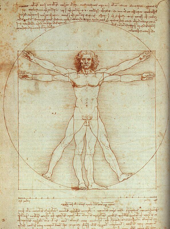
8. During the Renaissance, many great European universities were established.
(a) Andreas Vesalius and Leonardo da Vinci were renowned Renaissance men who produced monumental studies of the human form.
(b) De Humani Corporis Fabrica, written by Vesalius, had a tremendous impact on the advancement of human anatomy. Vesalius is regarded as the father of human anatomy.
9. Two major scientific contributions of the seventeenth and eighteenth centuries were the explanation of blood flow and the development of the microscope.
(a) In 1628, William Harvey correctly described the circulation of blood.
(b) Shortly after the microscope had been perfected by Antoni van Leeuwenhoek, many investigators added new discoveries to the rapidly changing specialty of microscopic anatomy.
10. The cell theory was formulated during the nineteenth century by Matthias Schleiden and Theodor Schwann, and cellular biology became established as a science separate from anatomy.
11. A trend toward simplification and standardization of anatomical nomenclature began in the twentieth century. In addition, many specialties within anatomy developed, including cytology, histology, embryology, electron microscopy, and radiology.
Danil Hammoudi.MD
Sinoe Medical Association

|
For those with slow connection click here |
To understand the abnormal you have to know the normal.
"FAILURE IS NOT AN OPTION"
Never Give Up
Yes it is hard, yes you will not sleep, yes it is a lot of material for a short period of time, but A is possible.
Hello Students of PGC AND LAUREL
In this site you will find all we talked about in Lab and more.
It will be updated after each Lab and before each lectures, question will be put on the site after each recitation for you to review.
Remember you will need to be ready before each lab and review after each lab.
The labs exercise and content can be downloaded at this address http://academic.pgcc.edu/AandP/ but it better to have your booklet.
)
If you have any question you get my E-mail
|
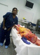
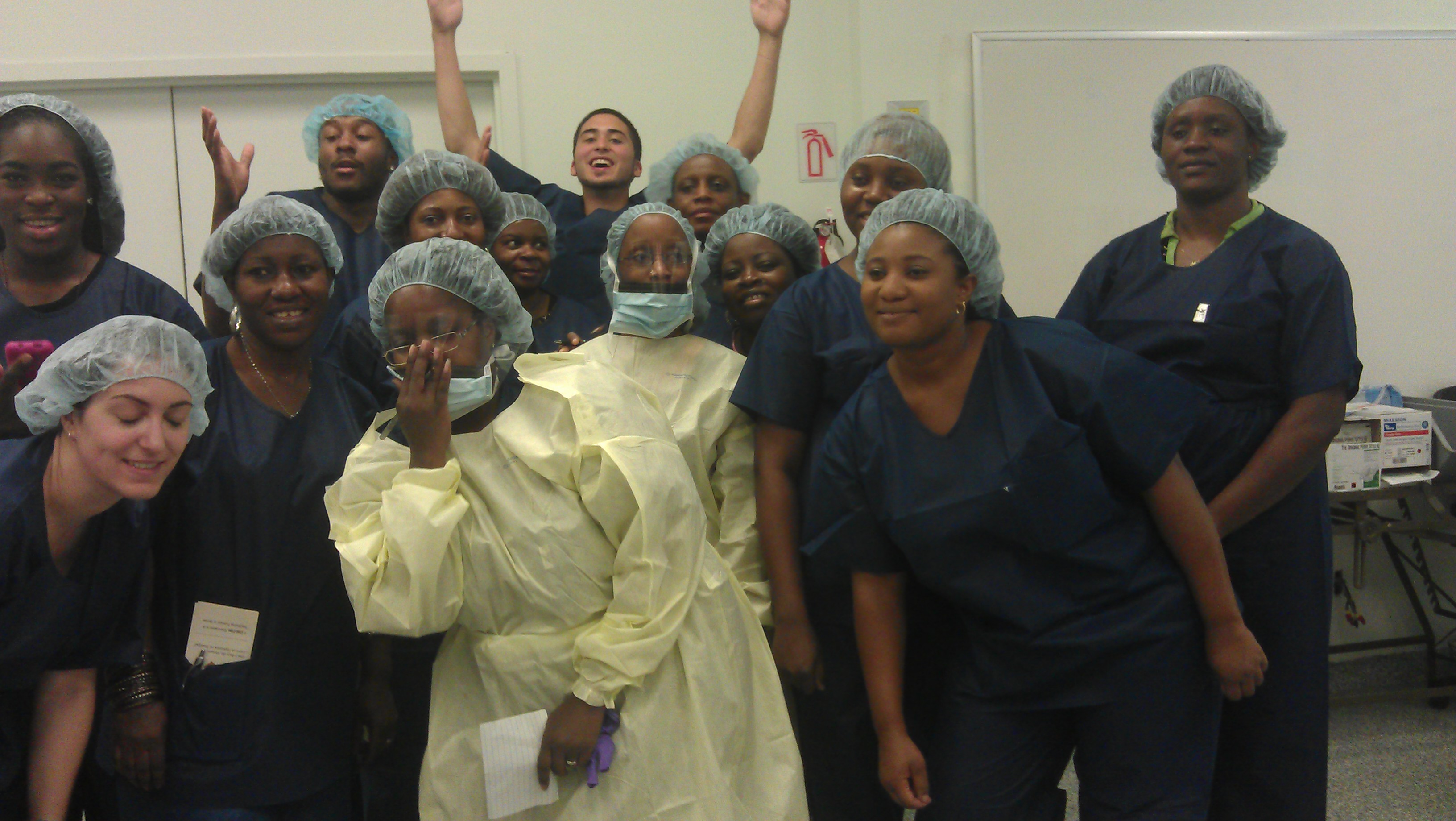
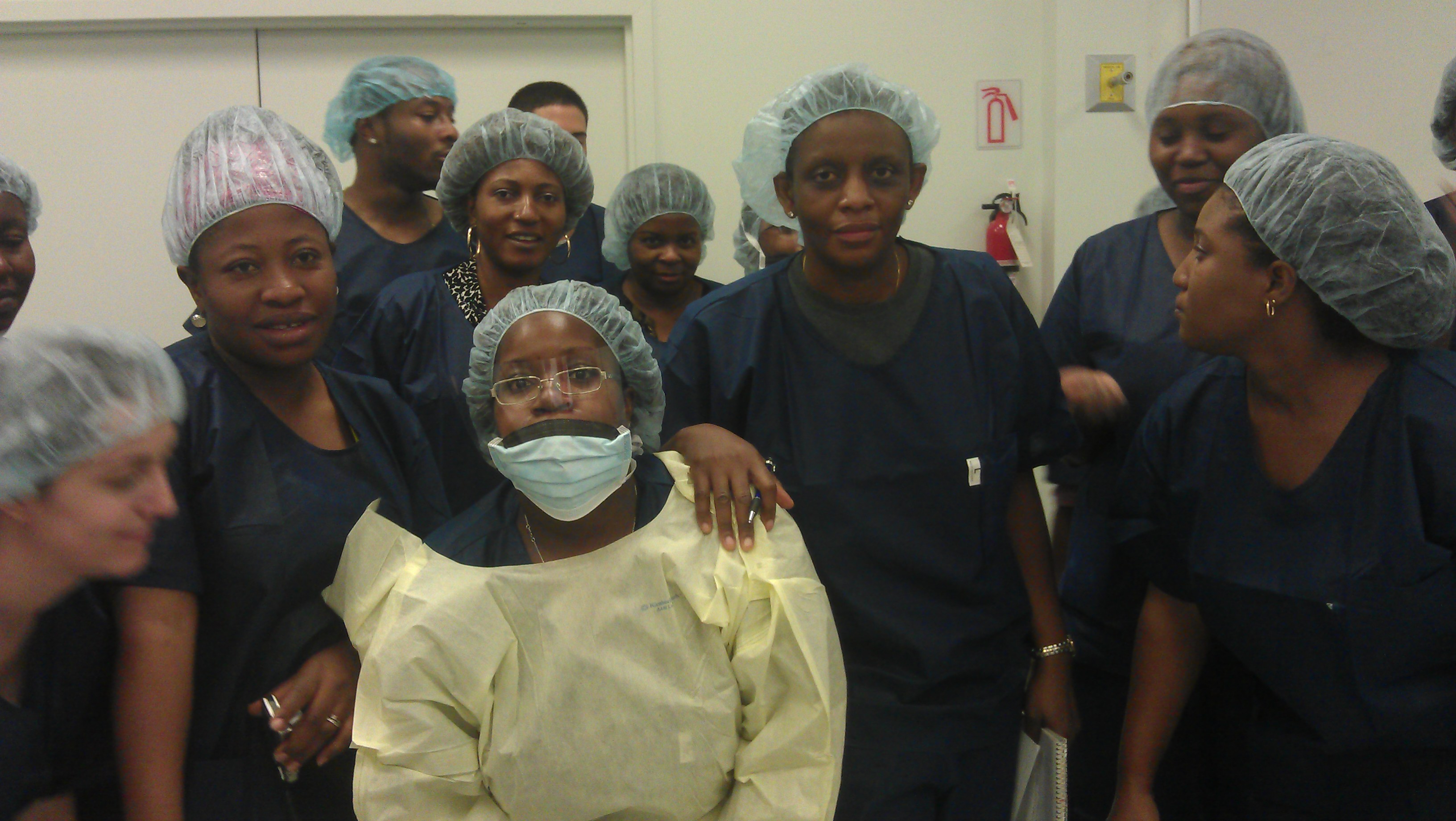
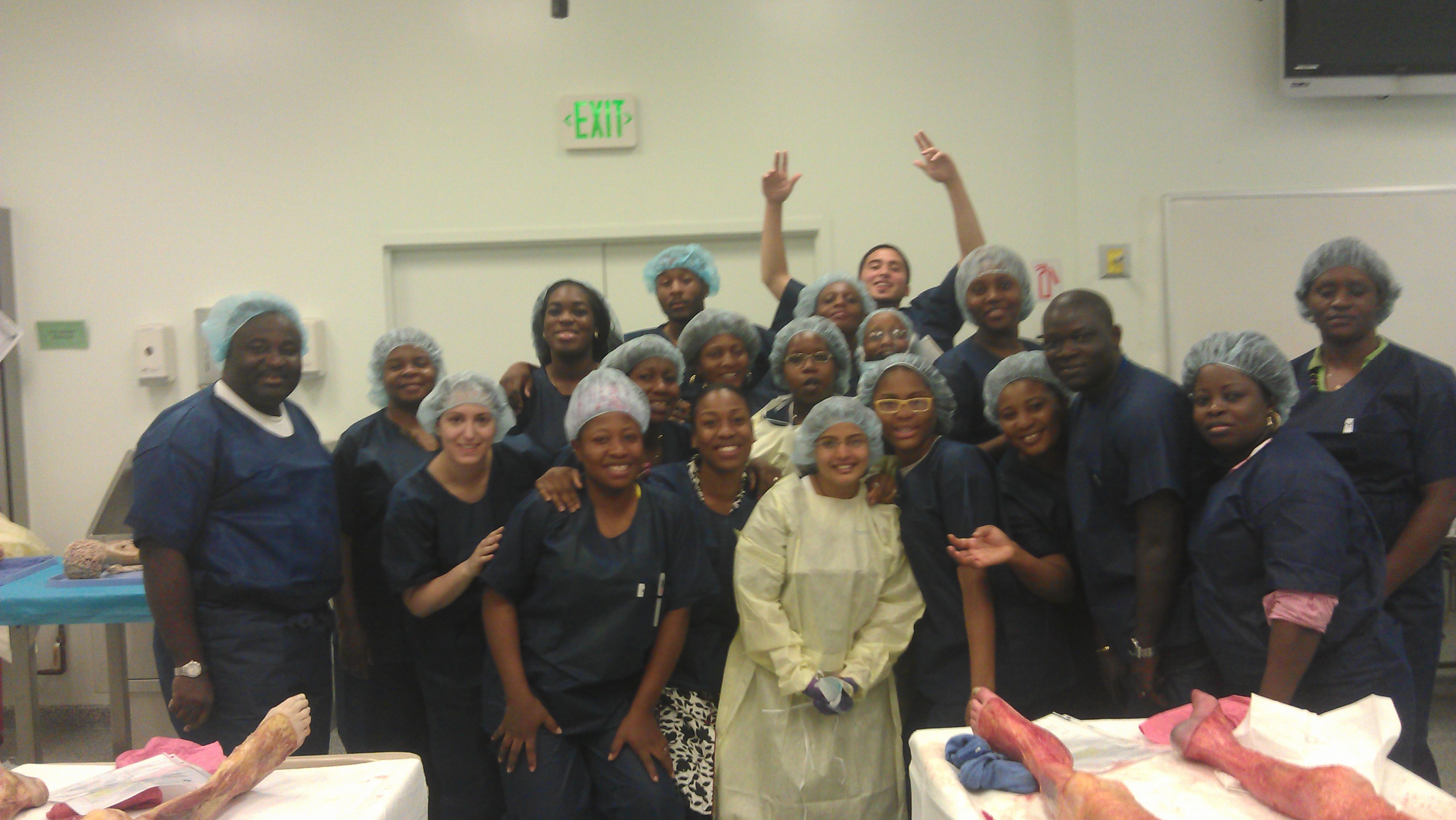
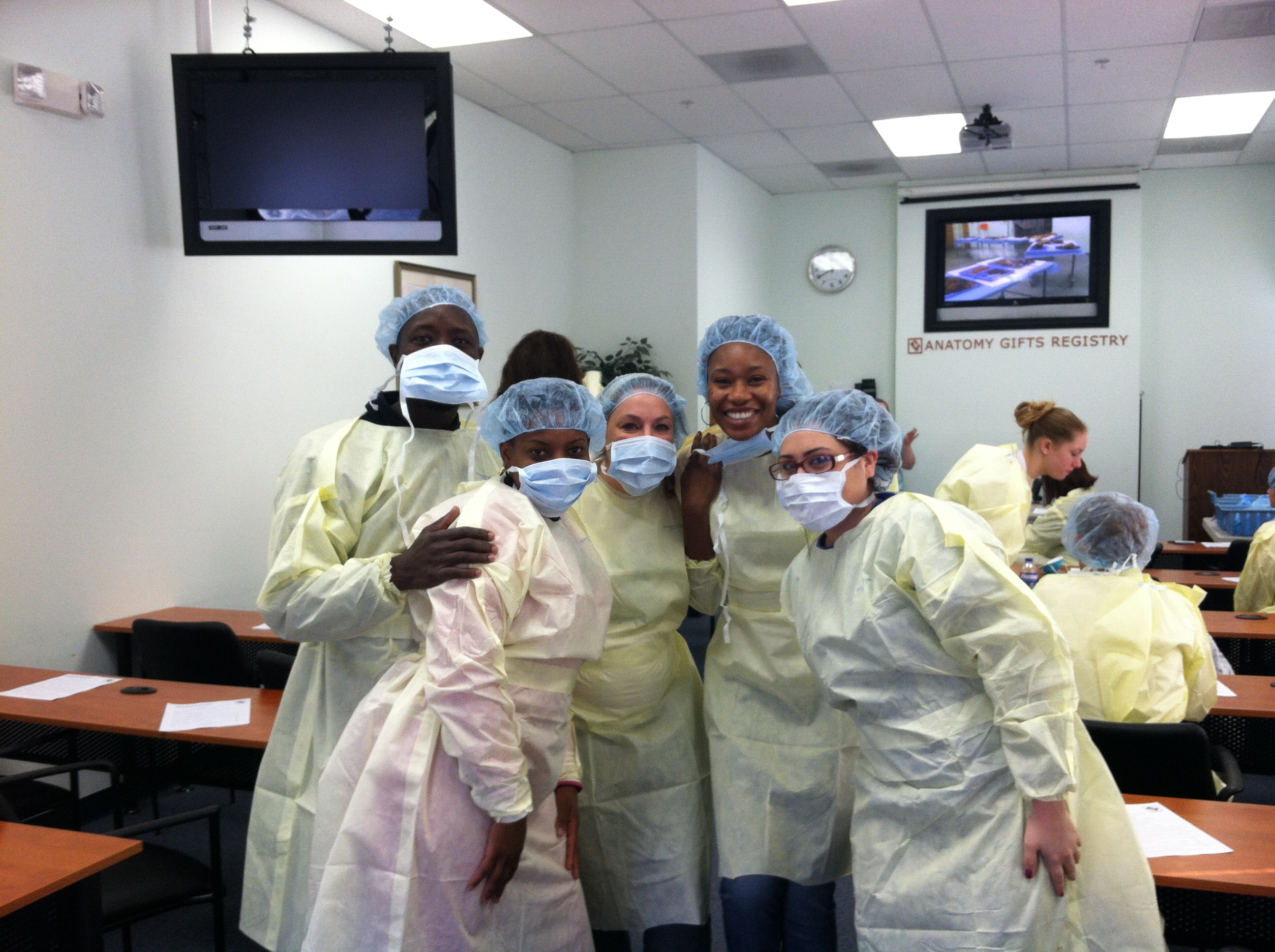
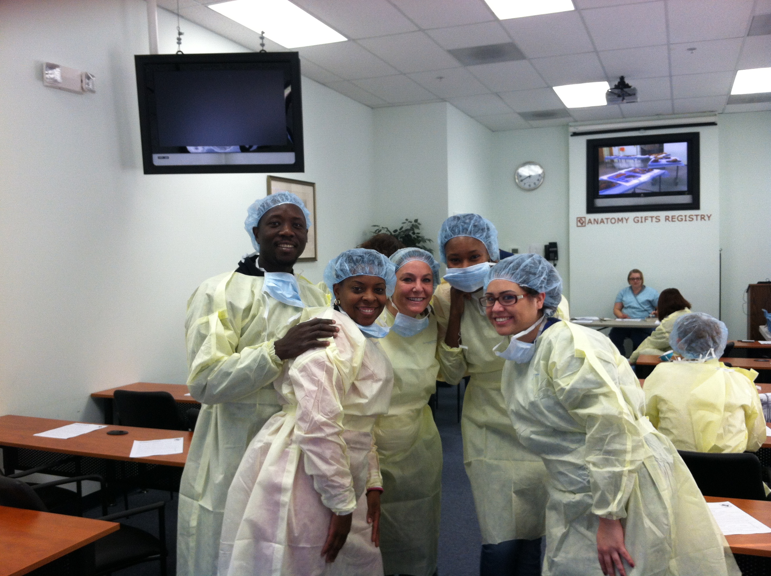
Dissection session for : http://anatomylab.anatomicgift.com/ Anatomy Gifts
Registry STUDENTS PICTURE FOR THE WET LAB
|

1. Human anatomy is the science concern with the structure of the human body.
2. The terms of anatomy are descriptive and are generally of Greek or Latin derivation.
3. The history of human anatomy parallels that of medicine and has also been greatly influenced by various religions.
|
1/ Generality and definition [txt]
2/ The cells [txt] 3/ The tissues [txt]
5/ Bones and Skeletal Tissues [txt]
6/ Nervous system introduction, histology
2/Body Regions and Major Body Cavities 8/Pelvis and lower extremities
1/ Epithelium and Connective Tissue 6/Neurophysiology [use your book CD] 7/ Muscles Generality and histology
|
All practical question are password protected now. due to abuse and copyright, my students only will have the pass
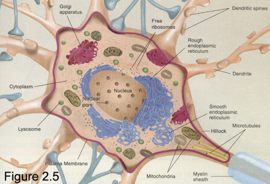
The cell is the structural and functional unit
of all living organisms, and is sometimes called the
"building block of life."Some organisms, such as
bacteria, are unicellular, consisting of a single cell.
Other organisms, such as humans, are multicellular.
(Humans have an estimated 100 trillion or 1014
cells; a typical cell size is 10 µm; a typical cell
mass is 1 nanogram.) The largest known cell is an
ostrich egg. The cell theory, first developed in 1839 by Schleiden and Schwann, states that all organisms are composed of one or more cells. All cells come from preexisting cells. Vital functions of an organism occur within cells, and all cells contain the hereditary information necessary for regulating cell functions and for transmitting information to the next generation of cells. The word cell comes from the Latin cellula, a small room. The name was chosen by Robert Hooke when he compared the cork cells he saw to the small rooms monks lived in. [http://en.wikipedia.org/wiki/Cell_(biology) ]
|


|
Anatomy (ənăt'əmē) , branch of biology concerned with the study of body structure of various organisms, including humans. Comparative anatomy is concerned with the structural differences of plant and animal forms.
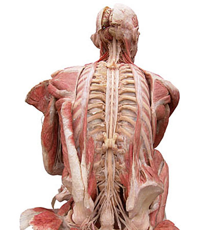
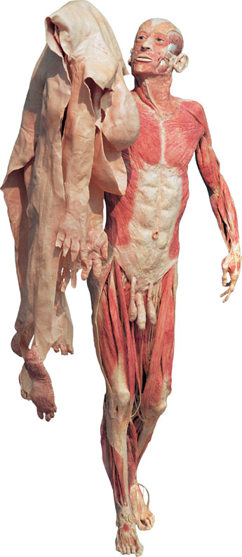
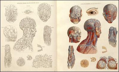
Gunther von Hagens, the German anatomist who created "Body Worlds," poses with one of his displays. The exhibit puts real human specimens on show, such as this one in Dallas. (Media Credit: Associated Press)Christopher Placek The study of similarities and differences in anatomical structures forms the basis for classification of both plants and animals. Embryology (see embryo) deals with developing plants or animals until hatching or birth (or germination, in plants); cell biology covers the internal anatomy of the cell, while histology is concerned with the study of aggregates of similarly specialized cells, called tissues. Related to anatomy is morphology, which involves comparative study of the corresponding organs in humans and animals. There are four major types of tissue present in the human body: epithelial tissue (see epithelium), muscular tissue (see muscle), connective tissue, and nervous tissue (see nervous system). Human anatomy is often studied by considering the individual systems that are composed of groups of tissues and organs; such systems include the skeletal system (see skeleton), muscular system, cutaneous system (see skin), circulatory system (including the lymphatic system), respiratory system (see respiration), digestive system, reproductive system, urinary system, and endocrine system. Little was known about human anatomy in ancient times because dissection, even of corpses, was widely forbidden. In the 2d cent., Galen, largely on the basis of animal dissection, made valuable contributions to the field. His work remained authoritative until the 14th and 15th cent., when a limited number of cadavers were made available to the medical schools. A better understanding of the science was soon reflected in the discoveries of Vesalius, William Harvey, and John Hunter. Various modern technologies have significantly refined the study of anatomy: X rays, CAT scans, and magnetic resonance imaging (MRI) are only several of the tools used today to obtain clear, accurate representations of the inner human anatomy. In 1994, for the first time, a detailed three-dimensional map of an entire human being (an executed prisoner who volunteered his body) was made available worldwide via the Internet using data from thousands of photographs, CAT scans, and MRIs of tiny cross sections of the body.[H. Gray, Gray's Anatomy (1987).]
|
| Longitudinal (interhemispheric) fissure | Between the cerebral hemispheres. Its floor is the corpus callosum. | |
| Lateral sulcus (sylvian fissure) | Separates temporal lobe from frontal and parietal lobes | |
| Insula | Landmark for underlying lentiform nucleus | |
| Central sulcus | Landmark for primary motor and somatic sensory areas | |
| Frontal lobe | ||
| Precentral gyrus | Primary motor area | |
| Inferior frontal gyrus | Includes Broca's expressive speech area | |
| Prefrontal cortex | Complex functions involving foresight | |
| Anterior cingulate cortex | Memory; movement | |
| Premotor area | Control of movement | |
| Supplementary motor area | Initiation of movements | |
| Frontal eye field | Saccadic eye movements (not pursuit or vergence) | |
| Parietal lobe | ||
| Postcentral gyrus | Primary somatosensory area | |
| Superior parietal cortex | Somatosensory association area; lesions cause apraxia, neglect | |
| Inferior parietal cortex | Higher order association areas for language, calculation; lesions cause receptive aphasia | |
| Posterior parietal cortex | Higher order association area for vision; eye-field for pursuit movements | |
| Occipital lobe | ||
| Calcarine
sulcus
(and the adjacent gyri) |
Primary visual area | |
| Remainder of the occipital lobe | Visual association cortex | |
| Temporal lobe | ||
| Inferior
and
inferolateral regions |
Highest order visual association area, including memories of complex scenes | |
| Superior temporal gyrus: | ||
| Anterior
&
middle part of superior surface |
Primary auditory area | |
| Middle and posterior parts | Auditory association area; also called Wernicke's area; part of the association area for language | |
| Parahippocampal gyrus: | ||
| Uncus | Primary olfactory cortex | |
| Entorhinal area | Includes primary and association cortex for olfaction. Afferents from all sensory association areas. Efferents to the hippocampus. | |
| Fusiform
gyrus
(lateral to collateral sulcus) |
Includes visual association cortex for remembering people's faces | |
| Hippocampal
formation
(subiculum, hippocampus, dentate gyrus etc) |
Integration of sensory experiences and formation of memories | |
| Medial surface of hemisphere | ||
| The
cingulate
gyrus, cingulate sulcus and parieto-occipital sulcus are conspicuous landmarks. |
The corpus callosum interconnects symmetrical areas of cerebral cortex of the two cerebral hemispheres |
|
Material links review |
|
http://academic.pgcc.edu/AandP/ http://academic.pg.cc.md.us/~mhubley/a&p/205/labimages.htm Atlas of Human Anatomy in Cross SectionMark J. Hubley, PhD http://academic.pgcc.edu/~mhubley/
|
Human Anatomy Books from Elsevier Health Information
and
diagrams of the human body systems covered in
A&P I & II http://www.innerbody.com/htm/body.html Digital
library
of health information and provides views of human
body structures and
systems covered in A&P I &
II http://www.thinkquest.org/library/cat_show.html?cat_id=129 https://histo.life.uiuc.edu/histo/ Histology
slides
of the human body structures covered in A&PI &
II http://www.uh.edu/sibs/tutorial/ap1.htm A&PI
tutoring
links http://www.MedicineNet.com http://www.healthsquare.com/ Contains
an
encyclopedia and medical
dictionary http://www.nlm.nih.gov/medlineplus A
medical dictionary,
medical encyclopedia
as well as
information on drugs and current health topics http://www.ama-assn.org/ama/pub/category/7140.html Medical
illustrations
of the human body http://www.vh.org/adult/provider/anatomy/HumanAnatomy/CrossSectionAtlas.html High-quality
photographs
of cross-sectional anatomy of the human
body http://www.meddean.luc.edu/lumen/ http://www.uofs.edu/sheep/human/frames1.html http://faculty.washington.edu/chudler/introb.html Exploration
of
the brain and the spinal cord
http://health.howstuffworks.com/ Lots
of
questions about health issues, and normal and abnormal
anatomy and physiology
of the human body An
article
explaining how the brain allows seeing,
hearing and
smelling the world http://faculty.washington.edu/chudler/eyecross.html
A
crossword puzzle on the anatomy and function of
the eye http://www.exploratorium.edu/memory/index.html Pictures
of
the dissected brain, as well as memory
games and tips for
improving memory http://www.pbs.org/wgbh/nova/coma/geography/ A
map of the human brain http://www.xecu.net/kiirenza/anatomy/ Anatomy
lecture
notes, as well as slides and photos of models and
dissected structures
of the human body http://faculty.washington.edu/chudler/auto.html The
sympathetic
and parasympathetic divisions of the autonomic
nervous system http://www.rcs.rome.ga.us/hargett/anatomy/nervous/index.htm A
review of the nervous system Many
links
to news on neurological disorders and
stroke http://www.emc.Maricopa.edu/faculty/farabee/BIOBK/BioBookENDOCR.html Components
and
functions of the endocrine system, and its
interactions with the
nervous system References
on
many disorders and diseases http://www.AmericanDiabeticAssociation.com Pathology
of
diabetes and its effects on the human body http://rarediseases.about.com http://www.mhhe.com/biosci/ap/holeessentials/student/olc/index.htm Case
studies
and tests on human body systems, and a review of General Biology http://cwx.prenhall.com/bookbind/pubbooks/martini6 http://library.thinkquest.org/TQ0312238/cgi-bin/view.cgi Explores
the brain National
Institute
of Health |